حدة البصر
| حدة البصر | |
|---|---|
| التشخيص الطبي | |
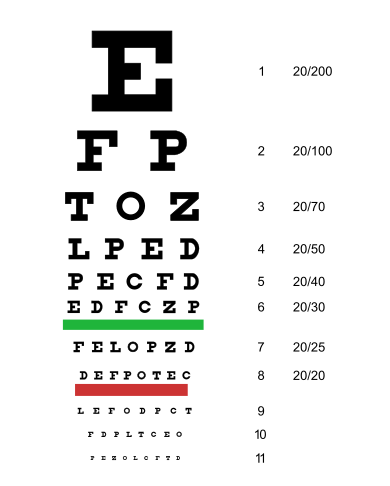
 عادة ما يستخدم جدول سنلن لاختبار حدة البصر. | |
| MeSH | D014792 |
| MedlinePlus | 003396 |
| LOINC | 28631-0 |
حدة البصر (Visual acuity، اختصاراً VA)، هو مصطلح عادة ما يشير إلى وضوح الرؤية، لكنه من الناحية التقنية يصنف قدرة الحيوان أو الإنسان على التعرف على التفاصيل الصغيرة بدقة. تعتمد حدة البصر على العوامل البصرية والعصبية. تؤثر العوامل البصرية للعين على حدة الصورة على الشبكية. تشمل العوامل العصبية صحة ووظيفة شبكية العين، والمسارات العصبية إلى المخ، والقدرة التفسيرية للمخ.[1]
إن حدة البصر التي يشار إليها عادةً هي "حدة البصر عن بعد" أو "وضوح الرؤية عن بعد" (على سبيل المثال، "نظر 20/20" أو "نظر 6/6)، والتي تصف قدرة الشخص على التعرف على التفاصيل الصغيرة من على مسافة بعيدة. هذه القدرة ضعيفة لدى الأشخاص المصابين بقصر النظر. هناك حدة بصرية أخرى هي "الحدة القريبة"، والتي تصف قدرة الشخص على التعرف على التفاصيل الصغيرة على مسافة قريبة. هذه القدرة ضعيفة لدى الأشخاص المصابين ببُعد النظر، والمعروف أيضاً باسم مد النظر.
من الأسباب البصرية الشائعة لضعف حدة البصر هو الخطا الانكساري: أخطاء في كيفية انكسار الضوء في العين. تشمل أسباب الأخطاء الانكسارية الانحرافات في شكل العين أو القرنية، وانخفاض قدرة العدسة على تركيز الضوء. عندما تكون القوة الانكسارية المجمعة للقرنية والعدسة عالية جدًا بالنسبة لطول العين، ستكون الصورة الشبكية في بؤرة التركيز أمام الشبكية وخارج التركيز على الشبكية، مما يؤدي إلى قصر النظر. تحدث صورة شبكية ضعيفة التركيز مماثلة عندما تكون القوة الانكسارية المجمعة للقرنية والعدسة منخفضة جدًا بالنسبة لطول العين باستثناء أن الصورة المركزة تكون خلف الشبكية، مما يؤدي إلى طول النظر. يشار إلى القوة الانكسارية الطبيعية باسم سواء البصر. تشمل الأسباب البصرية الأخرى لانخفاض حدة البصر اللابؤرية، حيث تكون خطوط اتجاه معين غير واضحة، ومخالفات القرنية الأكثر تعقيدًا.
يمكن تصحيح الأخطاء الانكسارية في الغالب باستخدام الوسائل البصرية (مثل النظارات الطبية، والعدسات اللاصقة، والجراحة الانكسارية). على سبيل المثال، في حالة قصر النظر، يكون التصحيح عن طريق تقليل قوة انكسار العين بواسطة ما يسمى بالعدسة السالبة.
توجد العوامل العصبية التي تحد من حدة البصر في الشبكية، أو في المسارات المؤدية إلى المخ، أو في المخ. ومن أمثلة الحالات التي تؤثر على الشبكية انفصال الشبكية والتنكس البقعي. ومن أمثلة الحالات التي تؤثر على المخ الغمش (الناجم عن عدم تطور المخ البصري بشكل صحيح في مرحلة الطفولة المبكرة) وتلف المخ، مثل الإصابة الدماغية الرضحية أو السكتة الدماغية. وعند تصحيح العوامل البصرية، يمكن اعتبار حدة البصر مقياسًا للأداء العصبي.
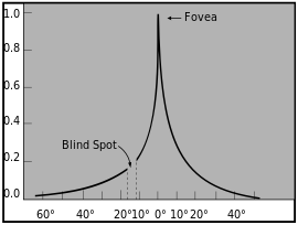
تقاس حدة البصر عادة أثناء التثبيت، أي كمقياس للرؤية المركزية (أو النقرة)، وذلك لأنها أعلى في المركز تمامًا.[2][3] ومع ذلك، فإن حدة البصر في الرؤية المحيطية قد تكون لها نفس الأهمية في الحياة اليومية. تتراجع حدة البصر نحو المحيط بشكل حاد أولاً ثم بشكل تدريجي، بطريقة خطية عكسية (أي أن الانحدار يتبع تقريبًا قطع زائد).[4][5] يكون الانحدار بحسب E2/(E2+E)، حيث E هو الانحراف في درجات الزاوية البصرية، وE2 هو ثابت يبلغ تقريبًا درجتين.[4][6][7] على سبيل المثال، عند درجتين من الانحراف، تكون حدة البصر نصف القيمة البقعية.
حدة البصر هي مقياس لمدى دقة حل التفاصيل الصغيرة في مركز المجال البصري؛ وبالتالي فهي لا تشير إلى كيفية التعرف على الأنماط الأكبر. وبالتالي فإن حدة البصر وحدها لا يمكنها تحديد الجودة العامة للوظيفة البصرية.[8]
التعريف

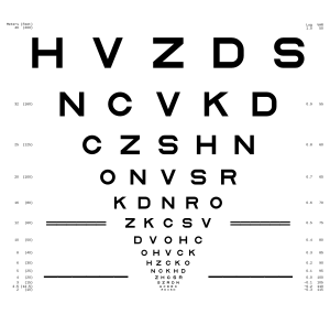
حدة البصر هي مقياس للدقة المكانية لنظام المعالجة البصرية. تُختبر حدة البصر، كما يشار إليها أحياناً من قبل المتخصصين في البصريات، من خلال مطالبة الشخص الذي يخضع لاختبار بصره بتحديد ما يسمى بالأنماط البصرية - الحروف المنمقة، حلقات لاندولت، رموز الأطفال، رموز للأميين، الحروف السيريلية الموحدة في جدول گولوڤين-سيڤتسڤ، أو أنماط أخرى - على مخطط مطبوع (أو بعض الوسائل الأخرى) من مسافة رؤية محددة. يتم تمثيل الأنماط البصرية كرموز سوداء على خلفية بيضاء (أي عند أقصى حد للتباين). تُضبط المسافة بين عيني الشخص ومخطط الاختبار بحيث تقترب من "اللانهاية البصرية" بالطريقة التي تحاول بها العدسة التركيز (حدة الرؤية البعيدة)، أو على مسافة قراءة محددة (حدة الرؤية القريبة).
القيمة المرجعية التي تعتبر حدة البصر فوقها طبيعية تسمى نظر 6/6 أو رؤية 6/6، والتي تعادلها رؤية 20/20 USC: على مسافة 6 أمتار أو 20 قدماً، تكون العين البشرية بهذا الأداء قادرة على فصل الخطوط التي تبعد عن بعضها البعض بمسافة 1.75 مم تقريباً.[9] تتوافق رؤية 6/12 تتوافق مع أداء أقل، في حين أن رؤية 6/3 تتوافق مع أداء أفضل. يتمتع الأفراد العاديون بحدة نظر تبلغ 6/4 أو أفضل (حسب العمر وعوامل أخرى).
في تعبير رؤية 6/x، يمثل البسط (6) المسافة بالأمتار بين الشخص والرسم البياني، ويمثل المقام (x) المسافة التي يستطيع عندها الشخص ذو حدة البصر 6/6 تمييز نفس النمط البصري. وبالتالي، فإن 6/12 يعني أن الشخص ذو الرؤية 6/6 يستطيع تمييز نفس النمط البصري من مسافة 12 متاًا (أي على ضعف المسافة). وهذا يعادل القول بأن الشخص ذو الرؤية 6/12 يمتلك نصف الدقة المكانية ويحتاج إلى ضعف الحجم لتمييز النمط البصري.
الطريقة البسيطة والفعّالة لتحديد حدة البصر هي تحويل الكسر إلى عدد عشري: 6/6 يتوافق إذن مع حدة البصر (أو الرؤية) 1.0 (انظر التعبير أدناه)، بينما يتوافق 6/3 مع 2.0، وهو ما يتحقق غالبًا لدى الشباب الأصحاء الذين تم تصحيح بصرهم جيدًا والذين يتمتعون برؤية ثنائية. تحديد حدة البصر كرقم عشري هو المعيار في البلدان الأوروپية، كما هو مطلوب من قبل المعيار الأوروپي (EN ISO 8596، سابقًا DIN 58220).
إن المسافة الدقيقة التي تقاس حدة البصر بها ليست مهمة طالما أنها بعيدة بما فيه الكفاية وحجم النمط البصري على الشبكية هو نفسه. يُحدد هذا الحجم كزاوية بصرية، وهي الزاوية عند العين التي يظهر النمط البصري تحتها. بالنسبة لـ 6/6 = 1.0 حدة بصرية، فإن حجم الحرف على جدول سنلن أو جدول لاندولت سي هو زاوية بصرية مقدارها 5 دقائق قوسية (1 دقيقة قوسية = 1/60 من الدرجة)، وهو خط مقاس 43 نقطة على بعد 20 قدماً.[10] من خلال تصميم نموذج بصري نموذجي (مثل مخطط سنلن E أو لاندولت C)، فإن الفجوة الحرجة التي يجب حلها هي 1/5 هذه القيمة، أي 1 قوس دقيقة. والأخيرة هي القيمة المستخدمة في التعريف الدولي لحِدة البصر:
acuity = 1/gap size [arc min].
إن حدة البصر هي مقياس للأداء البصري ولا ترتبط بوصفة النظارات المطلوبة لتصحيح الرؤية. وبدلاً من ذلك، يسعى فحص العين إلى إيجاد الوصفة التي ستوفر أفضل أداء بصري مصحح يمكن تحقيقه. وقد تكون حدة البصر الناتجة أكبر أو أقل من 6/6 = 1.0. والواقع أن الشخص الذي تم تشخيصه بأنه لديه رؤية 6/6 غالبًا ما يكون لديه حدة بصرية أعلى لأنه بمجرد الوصول إلى هذا المعيار، يُعتبر الشخص لديه رؤية طبيعية (بمعنى غير مضطربة) ولا يتم اختبار الأنماط البصرية الأصغر. قد يستفيد الأشخاص الذين لديهم رؤية 6/6 أو "أفضل" (20/15، 20/10، إلخ) من تصحيح النظارات لمشاكل أخرى تتعلق بالجهاز البصري، مثل طول النظر، أو إصابات العين، أو طول النظر الشيخوخي.
القياس
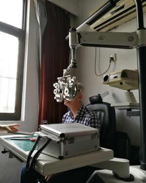
تقاس حدة البصر من خلال إجراء نفسي فيزيائي وبالتالي فهو يربط بين الخصائص الفيزيائية للمحفز وإدراك الشخص واستجاباته الناتجة. يمكن إجراء القياس باستخدام مخطط العين الذي اخترعه Ferdinand Monoyer، أو باستخدام أدوات بصرية، أو من خلال اختبارات محوسبة[11] مثل FrACT.[12]
ويجب الحرص على أن تتوافق ظروف المشاهدة مع المعايير،[13] مثل الإضاءة الصحيحة للغرفة ومخطط العين، ومسافة الرؤية الصحيحة، والوقت الكافي للاستجابة، وبدل الخطأ، وما إلى ذلك. في البلدان الأوروپية، يتم توحيد هذه الظروف من خلال المعيار الأوروپي (EN ISO 8596، سابقًا DIN 58220).
التاريخ
| السنة | الحدث |
|---|---|
| 1843 | اختراع أنواع اختبارات الرؤية عام 1843 بواسطة طبيب العيون الألماني هاينريش كوشلر (1811–1873)، في دارمشتات، ألمانيا. وقد جادل في الحاجة إلى توحيد اختبارات الرؤية وأنتج ثلاثة مخططات قراءة لتجنب الحفظ. |
| 1854 | إدوارد ياگر فون ياكستهال، وهو طبيب عيون من [ڤفيينا]]، يُجري تحسينات على أنواع اختبارات مخططات العين التي طورها هاينريش كويشلر. وينشر، باللغات الألمانية والفرنسية والإنگليزية ولغات أخرى، مجموعة من عينات القراءة لتوثيق الرؤية الوظيفية. ويستخدم الخطوط التي كانت متوفرة في دار الطباعة الحكومية في ڤيينا عام 1854 ويضع عليها علامات بالأرقام من كتالوج دار الطباعة تلك، والمعروفة حاليًا باسم أرقام ياگر. |
| 1862 | هرمان سنلن، طبيب عيون هولندي، ينشر في أوترخت كتابه [اختبار الحروف لقياس حدة البصر] Probebuchstaben zur Bestimmung der Sehschärfe ، والذي يحتوي على مخططات لقياس حدة البصر.[14]
في الإصدارات اللاحقة من كتابه، أطلق سنلن على حروف مخططاته اسم الأنماط البصرية ودعا إلى إجراء اختبارات رؤية موحدة.[15] لا تتطابق أنماط سنلن البصرية مع الحروف الاختبارية المستخدمة اليوم. فقد طُبعت بخط "پاراگون مصري" (أي باستخدام الخطوط المذيلة).[16][17] |
| 1888 | إدمون لاندولت يقدم الحلقة المفتوحة، المعروفة الآن باسم حلقة لاندولت، والتي أصبحت فيما بعد معياراً دولياً.[18][19] |
| 1894 |
تيودور ڤرتهايم يقدم في برلين قياسات تفصيلية لحدة البصر في الرؤية المحيطية.[4][20] |
| 1978 |
هيو تايلور يستخدم مبادئ التصميم هذه في "مخطط E المتساقط" للأميين، والذي تم استخدامه لاحقًا[21] لدراسة حدة البصر لدى الأستراليين الأصليين.[17] |
| 1982 | ريك فريس وآخرون من المعهد الوطني للعيون يختارون مخطط LogMAR، الذي تم تنفيذه باستخدام حروف سلون، لإنشاء طريقة موحدة لقياس حدة البصر لدراسة العلاج المبكر لاعتلال الشبكية السكري (ETDRS).
تُستخدم هذه المخططات في جميع الدراسات السريرية اللاحقة، وقد فعلت الكثير لتعريف المهنة بالمخطط الجديد والتقدم. تم استخدام البيانات من ETDRS لاختيار مجموعات الحروف التي تمنح كل سطر نفس الصعوبة المتوسطة، دون استخدام جميع الحروف في كل سطر. |
| 1984 | المجلس الدولي لطب العيون يعتمد "معيار جديد لقياس حدة البصر"، والذي يتضمن أيضاً الميزات المذكورة أعلاه. |
| 1988 | أنطونيو مدينا وبرادفورد هاولاند من معهد مساتشوستس للتكنولوجيا يطوران مخطط اختبار عين جديد باستخدام حروف تصبح غير مرئية مع انخفاض حدة البصر، بدلاً من أن تكون غير واضحة كما هو الحال في المخططات القياسية. لقد أظهرا الطبيعة التعسفية لكسر سنيلين وحذرا من دقة حدة البصر التي يتم تحديدها باستخدام مخططات من أنواع مختلفة من الحروف، والتي تم معايرتها بواسطة نظام سنلن.[22] |
الفسيولوجيا
الرؤية النهارية (أي الرؤية الضوئية) تخدمها خلايا مستقبلة مخروطية ذات كثافة مكانية عالية (في النقرة المركزية) وتسمح برؤية مرتفعة تبلغ 6/6 أو أفضل. في الضوء المنخفض (أي الرؤية الظلامية)، لا تتمتع المخاريط بحساسية كافية وتخدم القضبان الرؤية. وبالتالي تكون الدقة المكانية أقل بكثير. ويرجع هذا إلى الجمع المكاني القضبان، أي أن عددًا من القضبان يندمج في خلية ثنائية القطب، والتي تتصل بدورها بخلية عقدية، وتكون الوحدة الناتجة عن ذلك كبيرة، والحدة صغيرة. لا توجد قضبان في منتصف مجال الرؤية (النقرة)، ويتحقق أعلى أداء في الضوء المنخفض في الرؤية المحيطية القريبة.[4]
الدقة الزاوية القصوى للعين البشرية هي 28 ثانية قوسية أو 0.47 دقيقة قوسية؛[23] وهذا يعطي دقة زاوية قدرها 0.008 درجة، وعلى مسافة 1 كم يتوافق مع 136 مم. وهذا يساوي 0.94 دقيقة قوسية لكل زوج من الخطوط (خط أبيض وخط أسود)، أو 0.016 درجة. وبالنسبة لزوج من الپكسل (پكسل أبيض وپكسل أسود)، فإن هذا يعطي كثافة بكسل تبلغ 128 پكسل لكل درجة (PPD).
تُعرف الرؤية 6/6 على أنها القدرة على حل نقطتين من الضوء تفصل بينهما زاوية بصرية تبلغ دقيقة قوسية واحدة، وهو ما يعادل 60 نقطة في اليوم، أو حوالي 290-350 پكسل لكل بوصة لشاشة على جهاز محمول على مسافة 250-300 مم من العين.[24]
وبالتالي، فإن حدة البصر، أو القدرة على التمييز (في ضوء النهار، الرؤية المركزية)، هي خاصية المخاريط.[25] لحل التفاصيل، يجب على النظام البصري للعين أن يعرض صورة مركزة على النقرة، وهي منطقة داخل البقعة الصفراء تحتوي على أعلى كثافة من الخلايا المخروطية المستقبلة للضوء (النوع الوحيد من المستقبلات الضوئية الموجودة في مركز النقرة المركزية بقطر 300 ميكرومتر)، وبالتالي تتمتع بأعلى دقة وأفضل رؤية للألوان. على الرغم من أن حدة البصر ورؤية الألوان تتحكم فيهما نفس الخلايا، إلا أنهما وظيفتان فسيولوجيتان مختلفتان لا تترابطان إلا من خلال الموضع. يمكن أن تتأثر حدة البصر ورؤية الألوان بشكل مستقل.
إن حبيبات الفسيفساء الفوتوغرافية لها نفس القدرة المحدودة على التمييز مثل "حبيبات" الفسيفساء الشبكية. ولرؤية التفاصيل، يجب أن تتدخل مجموعة متوسطة من المستقبلات. والدقة القصوى هي 30 ثانية قوسية، والتي تتوافق مع قطر المخروط البقعي أو الزاوية التي تُحدد عند النقطة العقدية للعين. وللحصول على استقبال من كل مخروط، كما هو الحال إذا كانت الرؤية على أساس الفسيفساء، يجب الحصول على "الإشارة المحلية" من مخروط واحد عبر سلسلة من خلية ثنائية القطب وعقدية وركبية جانبية لكل منها. لكن العامل الرئيسي في الحصول على رؤية مفصلة هو التثبيط. ويتم ذلك بواسطة الخلايا العصبية مثل خلايا أماكرين والخلايا الأفقية، والتي تعمل على تعطيل انتشار الإشارات أو تقاربها. ويستمد هذا الميل إلى نقل الإشارات من واحد إلى واحد قوته من سطوع المركز ومحيطه، مما يؤدي إلى تحفيز التثبيط الذي يؤدي إلى توصيل واحد إلى واحد. ومع ذلك، فإن هذا السيناريو نادر، حيث قد تتصل المخاريط بالخلايا ثنائية القطب القزمة والمسطحة (المنتشرة)، ويمكن لخلايا أماكرين الأفقية دمج الرسائل بنفس سهولة تثبيطها.[9]
ينتقل الضوء من الجسم المثبت إلى النقرة عبر مسار وهمي يسمى المحور البصري. تؤثر أنسجة العين وهياكلها الموجودة في المحور البصري (وكذلك الأنسجة المجاورة له) على جودة الصورة. هذه الهياكل هي: الغشاء الدمعي، والقرنية، والحجرة الأمامية، والحدقة، والعدسة، والجسم الزجاجي، وأخيرًا الشبكية. الجزء الخلفي من الشبكية، المسمى الظهارة الصبغية الشبكية (RPE) مسؤول، من بين أشياء أخرى كثيرة، عن امتصاص الضوء الذي يعبر الشبكية حتى لا يتمكن من الارتداد إلى أجزاء أخرى من الشبكية. في العديد من الفقاريات، مثل القطط، حيث لا تشكل حدة البصر العالية أولوية، توجد طبقة من الغلاف العاكس تمنح المستقبلات الضوئية "فرصة ثانية" لامتصاص الضوء، وبالتالي تحسين القدرة على الرؤية في الظلام. وهذا هو ما يجعل عيون الحيوان تتوهج ظاهرياً في الظلام عندما يسلط عليها الضوء. كما أن للشبكة الشبكية وظيفة حيوية تتمثل في إعادة تدوير المواد الكيميائية التي تستخدمها القضبان والمخاريط في اكتشاف الفوتونات. وإذا تضررت الشبكة الشبكية ولم تنظف هذه الطبقة، فقد يؤدي ذلك إلى العمى.
كما هو الحال في عدسات التصوير الفوتوغرافي، تتأثر حدة البصر بحجم الحدقة. وتصل الانحرافات البصرية للعين التي تقلل من حدة البصر إلى أقصاها عندما تكون الحدقة في أكبر حجم لها (حوالي 8 مم)، وهو ما يحدث في ظروف الإضاءة المنخفضة. وعندما تكون الحدقة صغيرة (1-2 مم)، فقد تكون حدة الصورة محدودة بسبب حيود الضوء بواسطة الحدقة (انظر حد الحيود). وبين هذين الحدين يوجد قطر الحدقة الذي يكون أفضل بشكل عام لحدة البصر في العيون الطبيعية السليمة؛ ويميل هذا القطر إلى أن يكون حوالي 3 أو 4 مم.
إذا كانت بصريات العين مثالية من الناحية النظرية، فإن حدة البصر ستكون محدودة بحيود حدقة العين، والتي ستكون حدة محدودة بالحيود بمقدار 0.4 دقيقة قوسية أو رؤية 6/2.6. أصغر الخلايا المخروطية في الحفرة لها أحجام تتوافق مع 0.4 دقيقة قوسية من المجال البصري، مما يضع أيضاً حداً أدنى لحدة البصر. يمكن إثبات حدة البصر المثالية البالغة 0.4 دقيقة قوسية أو رؤية 6/2.6 باستخدام مقياس التداخل بالليزر الذي يتجاوز أي عيوب في بصريات العين ويسقط نمطاً من الأشرطة الداكنة والفاتحة مباشرة على الشبكية. تُستخدم مقاييس التداخل بالليزر الآن بشكل روتيني في المرضى الذين يعانون من مشاكل بصرية، مثل إعتام عدسة العين، لتقييم صحة الشبكية قبل إخضاعهم للجراحة.
القشرة البصرية هي جزء من القشرة المخية في الجزء الخلفي من المخ المسؤول عن معالجة المحفزات البصرية، ويسمى الفص القذالي. يمثل المجال المركزي 10 درجات (تقريباً امتداد البقعة الصفراء) ما لا يقل عن 60% من القشرة البصرية. ويعتقد أن العديد من هذه الخلايا العصبية تشارك بشكل مباشر في معالجة حدة البصر. يعتمد التطور السليم لحدة البصر الطبيعية على حصول الإنسان أو الحيوان على مدخلات بصرية طبيعية عندما يكون صغيراً جداً. أي حرمان من البصر، أي أي شيء يتداخل مع مثل هذه المدخلات على مدى فترة طويلة من الزمن، مثل إعتام عدسة العين، أو انحراف العين الشديد أو الحول، أو تفاوت الانكسار، أو تغطية العين أو تغطيتها أثناء العلاج الطبي، سيؤدي عادةً إلى انخفاض حاد ودائم في حدة البصر والتعرف على الأنماط في العين المصابة إذا لم يتم علاجها في وقت مبكر من الحياة، وهي الحالة المعروفة باسم الغمش. ينعكس انخفاض حدة البصر في العديد من التشوهات في خصائص الخلايا في القشرة البصرية. تتضمن هذه التغييرات انخفاضاً ملحوظاً في عدد الخلايا المتصلة بالعين المصابة وكذلك الخلايا المتصلة بكلتا العينين في المنطقة القشرية V1، مما يؤدي إلى فقدان الرؤية المجسمة، أي إدراك العمق من خلال الرؤية الثنائية (بالعامية: "الرؤية ثلاثية الأبعاد"). يشار إلى الفترة الزمنية التي يكون فيها الحيوان شديد الحساسية لمثل هذا الحرمان البصري بالفترة الحرجة.
ترتبط العين بالقشرة البصرية عن طريق العصب البصري الذي يخرج من مؤخرة العين. ويجتمع العصبان البصريان خلف العينين عند بالتصالب البصري، حيث يعبر حوالي نصف الألياف من كل عين إلى الجانب المقابل وينضمون إلى ألياف من العين الأخرى تمثل المجال البصري المقابل، وتشكل الألياف العصبية المدمجة من كلتا العينين السبيل البصري. ويشكل هذا في النهاية الأساس الفسيولوجي للرؤية الثنائية. وتمتد المسارات إلى محطة إعادة إرسال في الدماغ المتوسط تسمى النواة الركبية الوحشية، وهي جزء من المهاد، ثم إلى القشرة البصرية على طول مجموعة من الألياف العصبية تسمى الإشعاع البصري.
إن أي عملية مرضية في الجهاز البصري، حتى في البشر الأكبر سناً بعد الفترة الحرجة، غالباً ما تسبب انخفاضاً في حدة البصر. وبالتالي فإن قياس حدة البصر هو اختبار بسيط للوصول إلى صحة العينين أو المخ البصري أو المسار المؤدي إلى المخ. إن أي انخفاض مفاجئ نسبياً في حدة البصر هو دائماً مدعاة للقلق. الأسباب الشائعة لانخفاض حدة البصر هي إعتام عدسة العين والقرنية المتندبة، والتي تؤثر على المسار البصري، والأمراض التي تؤثر على شبكية العين، مثل التنكس البقعي ومرض السكري، والأمراض التي تؤثر على المسار البصري إلى المخ مثل الأورام والتصلب المتعدد، والأمراض التي تؤثر على القشرة البصرية مثل الأورام والسكتات الدماغية.
ورغم أن القدرة على التمييز تعتمد على حجم وكثافة المستقبلات الضوئية، فإن الجهاز العصبي لابد وأن يفسر معلومات المستقبلات. وكما تبين من التجارب التي أجريت على القطط والقردة، فإن الخلايا العقدية المختلفة في الشبكية مضبوطة على ترددات مكانية مختلفة، وبالتالي فإن بعض الخلايا العقدية في كل موقع تتمتع بحدة رؤية أفضل من غيرها. ولكن في نهاية المطاف، يبدو أن حجم رقعة من الأنسجة القشرية في القشرة البصرية V1 التي تعالج موقعاً معيناً في المجال البصري (وهو المفهوم المعروف باسم التكبير القشري) له نفس الأهمية في تحديد حدة البصر. وعلى وجه الخصوص، يكون هذا الحجم أكبر في مركز النقرة، ويقل مع زيادة المسافة من هناك.[4]
الجوانب البصرية
بالإضافة إلى الاتصالات العصبية للمستقبلات، فإن النظام البصري يلعب دوراً رئيسياً في دقة الشبكية. ففي العين المثالية، قد لا تتجاوز صورة محزز الحيود 0.5 ميكرومتر على الشبكية. لكن هذا ليس هو الحال بالتأكيد، علاوة على ذلك، يمكن أن تتسبب حدقة العين في حدوث حيود للضوء. وبالتالي، تختلط الخطوط السوداء على الشبكة بالخطوط البيضاء الفاصلة لتكوين مظهر رمادي. وقد تؤدي المشكلات البصرية المعيبة (مثل قصر النظر غير المصحح) إلى تفاقم الأمر، ولكن العدسات المناسبة يمكن أن تساعد. ويمكن شحذ الصور (مثل الشبكات) عن طريق التثبيط الجانبي، أي أن الخلايا الأكثر إثارة تمنع الخلايا الأقل إثارة. وينطبق نفس التفاعل في حالة الانحرافات اللونية، حيث يتم تثبيط الحواف الملونة حول الأشياء بالأبيض والأسود على نحو مماثل.[9]
التعبير
| 20 قدم | 10 قدم | 6 متر | 3 متر | العدد العشري | MAR | LogMAR |
|---|---|---|---|---|---|---|
| 20/1000 | 10/500 | 6/300 | 3/150 | 0.02 | 50 | 1.70 |
| 20/800 | 10/400 | 6/240 | 3/120 | 0.025 | 40 | 1.60 |
| 20/600 | 10/300 | 6/180 | 3/90 | 0.033 | 30 | 1.48 |
| 20/500 | 10/250 | 6/150 | 3/75 | 0.04 | 25 | 1.40 |
| 20/400 | 10/200 | 6/120 | 3/60 | 0.05 | 20 | 1.30 |
| 20/300 | 10/150 | 6/90 | 3/45 | 0.067 | 15 | 1.18 |
| 20/250 | 10/125 | 6/75 | 3/37 | 0.08 | 12.5 | 1.10 |
| 20/200 | 10/100 | 6/60 | 3/30 | 0.10 | 10 | 1.00 |
| 20/160 | 10/80 | 6/48 | 3/24 | 0.125 | 8 | 0.90 |
| 20/125 | 10/62 | 6/38 | 3/19 | 0.16 | 6.25 | 0.80 |
| 20/100 | 10/50 | 6/30 | 3/15 | 0.20 | 5 | 0.70 |
| 20/80 | 10/40 | 6/24 | 3/12 | 0.25 | 4 | 0.60 |
| 20/60 | 10/30 | 6/18 | 3/9 | 0.33 | 3 | 0.48 |
| 20/50 | 10/25 | 6/15 | 3/7.5 | 0.40 | 2.5 | 0.40 |
| 20/40 | 10/20 | 6/12 | 3/6 | 0.50 | 2 | 0.30 |
| 20/30 | 10/15 | 6/9 | 3/4.5 | 0.67 | 1.5 | 0.18 |
| 20/25 | 10/12 | 6/7.5 | 3/4 | 0.80 | 1.25 | 0.10 |
| 20/20 | 10/10 | 6/6 | 3/3 | 1.00 | 1 | 0.00 |
| 20/16 | 10/8 | 6/4.8 | 3/2.4 | 1.25 | 0.8 | −0.10 |
| 20/12.5 | 10/6 | 6/3.8 | 3/2 | 1.60 | 0.625 | −0.20 |
| 20/10 | 10/5 | 6/3 | 3/1.5 | 2.00 | 0.5 | −0.30 |
| 20/8 | 10/4 | 6/2.4 | 3/1.2 | 2.50 | 0.4 | −0.40 |
| 20/6.6 | 10/3.3 | 6/2 | 3/1 | 3.00 | 0.333 | −0.48 |
غالبًا ما تقاس حدة البصر وفقاً لحجم الحروف المعروضة على جدول سنلن أو حجم الرموز الأخرى، مثل لاندولت سي أو الجدول إي.
في بعض البلدان، يتم التعبير عن حدة البصر vulgar fraction، وفي بعضها كعدد عشري. باستخدام المتر كوحدة قياس، يتم التعبير عن حدة البصر (الكسرية) نسبة إلى 6/6. بخلاف ذلك، باستخدام القدم، يتم التعبير عن حدة البصر نسبة إلى 20/20. ولأغراض عملية، فإن الرؤية 20/20 تعادل 6/6. في النظام العشري، يتم تعريف حدة البصر على أنها القيمة العكسية لحجم الفجوة (مقاسة بالدقائق القوسية) لأصغر لاندولات سي، والتي يمكن تحديد اتجاهها بشكل موثوق. قيمة 1.0 تساوي 6/6.
LogMAR is another commonly used scale, expressed as the (decadic) logarithm of the minimum angle of resolution (MAR), which is the reciprocal of the acuity number. The LogMAR scale converts the geometric sequence of a traditional chart to a linear scale. It measures visual acuity loss: positive values indicate vision loss, while negative values denote normal or better visual acuity. This scale is commonly used clinically and in research because the lines are of equal length and so it forms a continuous scale with equally spaced intervals between points, unlike Snellen charts, which have different numbers of letters on each line.
A visual acuity of 6/6 is frequently described as meaning that a person can see detail from 6 metres (20 ft) away the same as a person with "normal" eyesight would see from 6 metres. If a person has a visual acuity of 6/12, they are said to see detail from 6 metres (20 ft) away the same as a person with "normal" eyesight would see it from 12 metres (39 ft) away.
The definition of 6/6 is somewhat arbitrary, since human eyes typically have higher acuity, as Tscherning writes, "We have found also that the best eyes have a visual acuity which approaches 2, and we can be almost certain that if, with a good illumination, the acuity is only equal to 1, the eye presents defects sufficiently pronounced to be easily established."[31] Most observers may have a binocular acuity superior to 6/6; the limit of acuity in the unaided human eye is around 6/3–6/2.4 (20/10–20/8), although 6/3 was the highest score recorded in a study of some US professional athletes.[32] Some birds of prey, such as hawks, are believed to have an acuity of around 20/2;[33] in this respect, their vision is much better than human eyesight.
When visual acuity is below the largest optotype on the chart, the reading distance is reduced until the patient can read it. Once the patient is able to read the chart, the letter size and test distance are noted. If the patient is unable to read the chart at any distance, they are tested as follows:
| الاسم | الاختصار | التعريف |
|---|---|---|
| Counting Fingers | CF | Ability to count fingers at a given distance. This test method is only used after it has been determined that the patient is not able to make out any of the letters, rings, or images on the acuity chart. The letters CF, and the testing distance, would represent the patient's acuity.
For example, the recording CF 5' would mean the patient was able to count the examiner's fingers from a maximum distance of 5 feet directly in front of the examiner. (The results of this test, on the same patient, may vary from examiner to examiner. This is due more to the size differences of the various examiners' hands and fingers, than to fluctuating vision.) |
| Hand Motion | HM | Ability to distinguish whether or not there is movement of the examiner's hand directly in front of the patient's eyes. This test method is only used after a patient shows little or no success with the Counting Fingers test. The letters HM, and the testing distance, would represent the patient's acuity.
For example, the recording HM 2' would mean that the patient was able to distinguish movement of the examiner's hand from a maximum distance of 2 feet directly in front of the examiner. (The results of the Hand Motion test are often recorded without the testing distance. This is due to the fact that this test is performed after the patient cannot "pass" the Counting Fingers test. At this point, the examiner is usually directly in front of the patient, and it is assumed that the Hand Motion test is performed at a testing distance of 1 foot or less.) |
| Light Perception | LP | Ability to perceive any light. This test method is used only after a patient shows little or no success with the Hand Motion test. In this test, an examiner shines a pen light at the patient's pupil and asks the patient to either point to the light source, or describe the direction that the light is coming from (up, out, straight ahead, down and out, etc.). If the patient is able to perceive light, the letters LP are recorded to represent the patient's acuity. If the patient is unable to perceive any light, the letters NLP (No Light Perception) are recorded. A patient with no light perception in one eye is considered blind in the respective eye. If NLP is recorded in both eyes, the patient is described as having total blindness. |
التعريفات القانونية
Various countries have defined statutory limits for poor visual acuity that qualifies as a disability. For example, in Australia, the Social Security Act defines blindness as:
A person meets the criteria for permanent blindness under section 95 of the Social Security Act if the corrected visual acuity is less than 6/60 on the Snellen Scale in both eyes or there is a combination of visual defects resulting in the same degree of permanent visual loss.[34]
In the US, the relevant federal statute defines blindness as follows:[35]
[T]he term "blindness" means central visual acuity of 20/200 or less in the better eye with the use of a correcting lens. An eye that is accompanied by a limitation in the fields of vision such that the widest diameter of the visual field subtends an angle no greater than 20 degrees shall be considered for purposes in this paragraph as having a central visual acuity of 20/200 or less.
A person's visual acuity is registered documenting the following: whether the test was for distant or near vision, the eye(s) evaluated and whether corrective lenses (i.e. glasses or contact lenses) were used:
- Distance from the chart
- D (distant) for the evaluation done at 20 feet (6 m).
- N (near) for the evaluation done at 15.7 inches (400 mm).
- Eye evaluated
- OD (Latin oculus dexter) for the right eye.
- OS (Latin oculus sinister) for the left eye.
- OU (Latin oculi uterque) for both eyes.
- Usage of spectacles during the test
- cc (Latin cum correctore) with correctors.
- sc: (Latin sine correctore) without correctors.
- Pinhole occluder
- The abbreviation PH is followed by the visual acuity as measured with a pinhole occluder, which temporarily corrects for refractive errors such as myopia or astigmatism.
- PHNI means no improvement of visual acuity using a pinhole occluder.
So, distant visual acuity of 6/10 and 6/8 with pinhole in the right eye will be: DscOD 6/10 PH 6/8. Distant visual acuity of count fingers and 6/17 with pinhole in the left eye will be: DscOS CF PH 6/17. Near visual acuity of 6/8 with pinhole remaining at 6/8 in both eyes with spectacles will be: NccOU 6/8 PH 6/8.
"Dynamic visual acuity" defines the ability of the eye to visually discern fine detail in a moving object.
اعتبارات القياس
Visual acuity measurement involves more than being able to see the optotypes. The patient should be cooperative, understand the optotypes, be able to communicate with the physician, and many more factors. If any of these factors is missing, then the measurement will not represent the patient's real visual acuity.
Visual acuity is a subjective test meaning that if the patient is unwilling or unable to cooperate, the test cannot be done. A patient who is sleepy, intoxicated, or has any disease that can alter their consciousness or mental status, may not achieve their maximum possible acuity.
Patients who are illiterate in the language whose letters and/or numbers appear on the chart will be registered as having very low visual acuity if this is not known. Some patients will not tell the examiner that they do not know the optotypes, unless asked directly about it. Brain damage can result in a patient not being able to recognize printed letters, or being unable to spell them.
A motor inability can make a person respond incorrectly to the optotype shown and negatively affect the visual acuity measurement.
Variables such as pupil size, background adaptation luminance, duration of presentation, type of optotype used, interaction effects from adjacent visual contours (or "crowding") can all affect visual acuity measurement.
الاختبار لدى الأطفال
The newborn's visual acuity is approximately 6/133, developing to 6/6 well after the age of six months in most children, according to a study published in 2009.[36]
The measurement of visual acuity in infants, pre-verbal children and special populations (for instance, disabled individuals) is not always possible with a letter chart. For these populations, specialised testing is necessary. As a basic examination step, one must check whether visual stimuli can be fixated, centered and followed.
More formal testing using preferential looking techniques use Teller acuity cards (presented by a technician from behind a window in the wall) to check whether the child is more visually attentive to a random presentation of vertical or horizontal gratings on one side compared with a blank page on the other side – the bars become progressively finer or closer together, and the endpoint is noted when the child in its adult carer's lap equally prefers the two sides.
Another popular technique is electro-physiologic testing using visual evoked (cortical) potentials (VEPs or VECPs), which can be used to estimate visual acuity in doubtful cases and expected severe vision loss cases like Leber's congenital amaurosis.
VEP testing of acuity is somewhat similar to preferential looking in using a series of black and white stripes (sine wave gratings) or checkerboard patterns (which produce larger responses than stripes). Behavioral responses are not required and brain waves created by the presentation of the patterns are recorded instead. The patterns become finer and finer until the evoked brain wave just disappears, which is considered to be the endpoint measure of visual acuity. In adults and older, verbal children capable of paying attention and following instructions, the endpoint provided by the VEP corresponds very well to the psychophysical measure in the standard measurement (i.e. the perceptual endpoint determined by asking the subject when they can no longer see the pattern). There is an assumption that this correspondence also applies to much younger children and infants, though this does not necessarily have to be the case. Studies do show the evoked brain waves, as well as derived acuities, are very adult-like by one year of age.
For reasons not totally understood, until a child is several years old, visual acuities from behavioral preferential looking techniques typically lag behind those determined using the VEP, a direct physiological measure of early visual processing in the brain. Possibly it takes longer for more complex behavioral and attentional responses, involving brain areas not directly involved in processing vision, to mature. Thus the visual brain may detect the presence of a finer pattern (reflected in the evoked brain wave), but the "behavioral brain" of a small child may not find it salient enough to pay special attention to.
A simple but less-used technique is checking oculomotor responses with an optokinetic nystagmus drum, where the subject is placed inside the drum and surrounded by rotating black and white stripes. This creates involuntary abrupt eye movements (nystagmus) as the brain attempts to track the moving stripes. There is a good correspondence between the optokinetic and usual eye-chart acuities in adults. A potentially serious problem with this technique is that the process is reflexive and mediated in the low-level brain stem, not in the visual cortex. Thus someone can have a normal optokinetic response and yet be cortically blind with no conscious visual sensation.
"حدة البصر الطبيعية"
Visual acuity depends upon how accurately light is focused on the retina, the integrity of the eye's neural elements, and the interpretative faculty of the brain.[37] "Normal" visual acuity (in central, i.e. foveal vision) is frequently considered to be what was defined by Herman Snellen as the ability to recognize an optotype when it subtended 5 minutes of arc, that is Snellen's chart 6/6-metre, 20/20 feet, 1.00 decimal or 0.0 logMAR. In young humans, the average visual acuity of a healthy, emmetropic eye (or ametropic eye with correction) is approximately 6/5 to 6/4, so it is inaccurate to refer to 6/6 visual acuity as "perfect" vision. On the contrary, Tscherning writes, "We have found also that the best eyes have a visual acuity which approaches 2, and we can be almost certain that if, with a good illumination, the acuity is only equal to 1, the eye presents defects sufficiently pronounced to be easily established."[31]
6/6 is the visual acuity needed to discriminate two contours separated by 1 arc minute – 1.75 mm at 6 metres. This is because a 6/6 letter, E for example, has three limbs and two spaces in between them, giving 5 different detailed areas. The ability to resolve this therefore requires 1/5 of the letter's total size, which in this case would be 1 minute of arc (visual angle). The significance of the 6/6 standard can best be thought of as the lower limit of normal, or as a screening cutoff. When used as a screening test, subjects that reach this level need no further investigation, even though the average visual acuity with a healthy visual system is typically better.
Some people may have other visual problems, such as severe visual field defects, color blindness, reduced contrast, mild amblyopia, cerebral visual impairments, inability to track fast-moving objects, or one of many other visual impairments and still have "normal" visual acuity. Thus, "normal" visual acuity does not imply normal vision. The reason visual acuity is very widely used is that it is easily measured, its reduction (after correction) often indicates some disturbance, and that it often corresponds with the normal daily activities a person can handle, and evaluates their impairment to do them (even though there is heavy debate over that relationship).
معايير أخرى
Normally, visual acuity refers to the ability to resolve two separated points or lines, but there are other measures of the ability of the visual system to discern spatial differences.
Vernier acuity measures the ability to align two line segments. Humans can do this with remarkable accuracy. This success is regarded as hyperacuity. Under optimal conditions of good illumination, high contrast, and long line segments, the limit to vernier acuity is about 8 arc seconds or 0.13 arc minutes, compared to about 0.6 arc minutes (6/4) for normal visual acuity or the 0.4 arc minute diameter of a foveal cone. Because the limit of vernier acuity is well below that imposed on regular visual acuity by the "retinal grain" or size of the foveal cones, it is thought to be a process of the visual cortex rather than the retina. Supporting this idea, vernier acuity seems to correspond very closely (and may have the same underlying mechanism) enabling one to discern very slight differences in the orientations of two lines, where orientation is known to be processed in the visual cortex.
The smallest detectable visual angle produced by a single fine dark line against a uniformly illuminated background is also much less than foveal cone size or regular visual acuity. In this case, under optimal conditions, the limit is about 0.5 arc seconds or only about 2% of the diameter of a foveal cone. This produces a contrast of about 1% with the illumination of surrounding cones. The mechanism of detection is the ability to detect such small differences in contrast or illumination, and does not depend on the angular width of the bar, which cannot be discerned. Thus as the line gets finer, it appears to get fainter but not thinner.
Stereoscopic acuity is the ability to detect differences in depth with the two eyes. For more complex targets, stereoacuity is similar to normal monocular visual acuity, or around 0.6–1.0 arc minutes, but for much simpler targets, such as vertical rods, may be as low as only 2 arc seconds. Although stereoacuity normally corresponds very well with monocular acuity, it may be very poor, or absent, even in subjects with normal monocular acuities. Such individuals typically have abnormal visual development when they are very young, such as an alternating strabismus, or eye turn, where both eyes rarely, or never, point in the same direction and therefore do not function together.
Another test, including Visual acuity (EVTS/OptimEyes), uses targets which change in size, contrast, and viewing time. This test was developed by Daniel M. Laby with colleagues and uses item response theory to calculate a vision performance score (core score). This specific test of visual function has been shown to correlate to professional sports performance.[38]
حدة الحركة
The eye has acuity limits for detecting motion.[39] Forward motion is limited by the subtended angular velocity detection threshold (SAVT), and horizontal and vertical motion acuity are limited by lateral motion thresholds. The lateral motion limit is generally below the looming motion limit, and for an object of a given size, lateral motion becomes the more insightful of the two, once the observer moves sufficiently far away from the path of travel. Below these thresholds subjective constancy is experienced in accordance with the Stevens' power law and Weber–Fechner law.
عتبة الكشف عن السرعة الزاوية المضمنة (SAVT)
There is a specific acuity limit in detecting an approaching object's looming motion.[40][41] This is regarded as the subtended angular velocity detection threshold (SAVT) limit of visual acuity.[42] It has a practical value of 0.0275 rad/s.[43] For a person with SAVT limit of , the looming motion of a directly approaching object of size S, moving at velocity v, is not detectable until its distance D is[40]
where the S2/4 term is omitted for small objects relative to great distances by small-angle approximation.
To exceed the SAVT, an object of size S moving as velocity v must be closer than D; beyond that distance, subjective constancy is experienced. The SAVT can be measured from the distance at which a looming object is first detected:
where the S2 term is omitted for small objects relative to great distances by small-angle approximation.
The SAVT has the same kind of importance to driving safety and sports as the static limit. The formula is derived from taking the derivative of the visual angle with respect to distance, and then multiplying by velocity to obtain the time rate of visual expansion (dθ/dt = dθ/dx · dx/dt).
الحركة الجانبية
There are acuity limits () of horizontal and vertical motion as well.[39] They can be measured and defined by the threshold detection of movement of an object traveling at distance D and velocity v orthogonal to the direction of view, from a set-back distance B with the formula
Because the tangent of the subtended angle is the ratio of the orthogonal distance to the set-back distance, the angular time rate (rad/s) of lateral motion is simply the derivative of the inverse tangent multiplied by the velocity (dθ/dt = dθ/dx · dx/dt). In application this means that an orthogonally traveling object will not be discernible as moving until it has reached the distance
where for lateral motion is generally ≥ 0.0087 rad/s with probable dependence on deviation from the fovia and movement orientation,[39] velocity is in terms of the distance units, and zero distance is straight ahead. Far object distances, close set-backs, and low velocities generally lower the salience of lateral motion. Detection with close or null set-back can be accomplished through the pure scale changes of looming motion.[41]
الحركة الشعاعية
The motion acuity limit affects radial motion in accordance to its definition, hence the ratio of the velocity v to the radius R must exceed :
Radial motion is encountered in clinical and research environments, in dome theaters, and in virtual-reality headsets.
انظر أيضاً
المصادر
- ^ Cline D, Hofstetter HW, Griffin J (1997). Dictionary of Visual Science (4th ed.). Boston: Butterworth-Heinemann. ISBN 978-0-7506-9895-5.
- ^ Acuity is highest in a tiny area, sometimes called the "foveal bouquet", with a diameter of only 8 – 16 minutes of arc (see Strasburger, 2020, p. 10)
- ^ Strasburger, H. (2020). "seven myths on crowding and peripheral vision". i-Perception. 11 (2): 1–45. doi:10.1177/2041669520913052. PMC 7238452. PMID 32489576.
- ^ أ ب ت ث ج ح Strasburger H, Rentschler I, Jüttner M (2011). "Peripheral vision and pattern recognition: a review". Journal of Vision. 11 (5): 13, 1–82. doi:10.1167/11.5.13. PMC 11073400. PMID 22207654.
{{cite journal}}: Check|pmc=value (help) - ^ Barghout-Stein L (1999). On differences between peripheral and foveal pattern masking (Thesis). University of California, Berkeley.
- ^ Anstis, S. M. (1974). "A chart demonstrating variations in acuity with retinal position". Vision Research. 14 (7): 589–592. doi:10.1016/0042-6989(74)90049-2. PMID 4419807.
- ^ Estimates of E2 vary quite a bit. The approximate value of 2 degrees is taken from Strasburger et al. (2011), Table 4. It results from Anstis's (1974) Figure 1, with the foveal value assumed to be the standard 20/20 acuity.
- ^ Kandel, Himal; Nguyen, Vuong; Piermarocchi, Stefano; Ceklic, Lala; Teo, Kelvin; Arnalich-Montiel, Francisco; Miotto, Stefania; Daien, Vincent; Gillies, Mark C.; Watson, Stephanie L. (2022). "Quality of life impact of eye diseases: a Save Sight Registries study". Clinical & Experimental Ophthalmology (in الإنجليزية). 50 (4): 386–397. doi:10.1111/ceo.14050. ISSN 1442-6404. PMC 9303885. PMID 35080803.
- ^ أ ب ت "eye, human". Encyclopædia Britannica 2006 Ultimate Reference Suite DVD. 2008.
- ^ Table 3, page 20, Morrison, James Barbour , Joanna Zander (April 2008). "Determining the appropriate font size, and use of colour and contrast for underwater displays - for Defence R & D Canada". researchgate. Retrieved 2023-12-20.
{{cite web}}: CS1 maint: multiple names: authors list (link) - ^ Strasburger H (2014). "Software for visual psychophysics: an overview". VisionScience.com.
- ^ Bach M (2016). "The Freiburg Visual Acuity Test".
- ^ Visual Functions Committee (25 May 1984). "Visual acuity measurement standard" (PDF). International Council of Ophthalmology. Archived from the original (PDF) on 7 May 2021. Retrieved 29 May 2015.
- ^ Snellen, Herman (1862). Probebuchstaben zur Bestimmung der Sehschärfe [Test letters for measuring visual acuity. Utrecht: P. W. Van De Weijer. Retrieved 12 September 2023.
- ^ Snellen, Herman (1885). Optotypi ad visum determinandum secundum formulam = d/D. Ed. 8, metrico systemate [ (8 ed.). London: Williams and Norgate. Retrieved 12 September 2023.
- ^ Enerson, Ole Daniel (2017). "Herman Snellen". Whonamedit?.
- ^ أ ب Colenbrander A (2001). "Measuring Vision and Vision Loss" (PDF). Archived from the original (PDF) on 4 December 2014.
- ^ Landolt E (1888). "Méthode optométrique simple" [A simple optometric method]. Bulletins et Mémoires de la Société Française d'Ophtalmologie (in الفرنسية) (6): 213–214.
- ^ Grimm; Rassow; Wesemann; Saur; Hilz (1994). "Correlation of optotypes with the Landolt Ring – a fresh look at the comparability of optotypes". Optometry and Vision Science. 71 (1): 6–13. doi:10.1097/00006324-199401000-00002. PMID 8146001. S2CID 24533843.
- ^ Wertheim T (1894). "Über die indirekte Sehschärfe" [On indirect visual acuity]. Zeitschrift für Psychologie und Physiologie der Sinnesorgane (in الألمانية) (7): 172–187.
- ^ Taylor H (1981). "Racial Variations in Vision". Am. J. Epidemiol. 113 (1): 62–80. doi:10.1093/oxfordjournals.aje.a113067. PMID 7457480.
- ^ Medina A, Howland B (1988). "A novel high-frequency visual acuity chart". Ophthalmic Physiol Opt. 8 (1): 14–8. doi:10.1016/0275-5408(88)90083-x. PMID 3419824.
- ^ Deering MF. "The Limits of Human Vision" (PDF)..
- ^ "Visual Acuity of the Human Eye". NDT Resource Center. Archived from the original on 6 September 2012. Retrieved 7 May 2006.
- ^ Ali MA, Klyne M (1985). Vision in Vertebrates. New York: Plenum Press. p. 28. ISBN 978-0-306-42065-8.
- ^ acuity as reciprocal of degrees visual angle, divided by the foveal value
- ^ Original figure in Østerberg, G. (1935). "Topography of the layer of rods and cones in the human retina". Acta Ophthalmologica. 13 (Suppl. 6): 11–103. doi:10.1111/j.1755-3768.1935.tb04723.x. S2CID 220560741.. Østerberg's figure is reproduced in Strasburger et al. (2011), Fig. 4
- ^ Hunziker H (2006). Im Auge des Lesers: foveale und periphere Wahrnehmung – vom Buchstabieren zur Lesefreude [The eye of the reader: foveal and peripheral perception – from letter recognition to the joy of reading] (in الألمانية). Zürich: Transmedia Stäubli Verlag. ISBN 978-3-7266-0068-6.
- ^ Rohrschneider, K. (2004). "Determination of the location of the fovea on the fundus". Investigative Ophthalmology & Visual Science. 45 (9): 3257–3258. doi:10.1167/iovs.03-1157. PMID 15326148.
- ^ "Contrast sensitivity" (PDF). LEA-Test Ltd. Retrieved 21 July 2018.
- ^ أ ب Physiologic Optics: Dioptrics of the Eye, Functions of the Retina, Ocular Movements and Binocular Vision
- ^ Kirschen DG, Laby DM (1 May 2006). "Sports Vision Testing: An Innovative Approach To Increase Revenues". Optometric Management.
- ^ Kirschbaum K. "Family Accipitridae". Animal Diversity Web. University of Michigan Museum of Zoology. Retrieved 30 January 2010.
- ^ Social Security Act 1991 (Cth) "Table 13, Schedule 1B". as at 20 September 2011.
- ^ 42 U.S.C. § 416(i)(1)(B) (Supp. IV 1986). Cited in "SSR 90-5c: Sections 216(i)(1)(B) and 223(c)(1) and (d)(1)(B) of the Social Security Act (42 U.S.C. 416(i)(1)(B) and 423(c)(1) and (d)(1)(B)) Disability insurance benefits – Interpreting the statutory blindness provision". 9 November 1990.
- ^ Pan Y, Tarczy-Hornoch K, Cotter SA (June 2009). "Visual acuity norms in pre-school children: the Multi-Ethnic Pediatric Eye Disease Study". Optom Vis Sci. 86 (6): 607–12. doi:10.1097/OPX.0b013e3181a76e55. PMC 2742505. PMID 19430325.
- ^ Carlson N, Kurtz D, Heath D, Hines C (1990). Clinical Procedures for Ocular Examination. Norwalk, CT: Appleton & Lange. ISBN 978-0-07-184920-3.
- ^ Laby, Daniel M.; Kirschen, David G.; Govindarajulu, Usha; DeLand, Paul (2019-11-14). "The Effect of Visual Function on the Batting Performance of Professional Baseball Players". Scientific Reports (in الإنجليزية). 9 (1): 16847. Bibcode:2019NatSR...916847L. doi:10.1038/s41598-019-52546-2. ISSN 2045-2322. PMC 6856529. PMID 31728011.
- ^ أ ب ت Lappin JS, Tadin D, Nyquist JB, Corn AL (January 2009). "Spatial and temporal limits of motion perception across variations in speed, eccentricity, and low vision". Journal of Vision. 9 (30): 30.1–14. doi:10.1167/9.1.30. PMID 19271900.
Displacement thresholds for peripheral motion were affected by acuity limits for speeds below 0.5 degrees/s. [0.0087 radians/s]
- ^ أ ب Weinberger H (19 February 1971). "Conjecture on the Visual Estimation of Relative Radial Motion". Nature. 229 (5286): 562. Bibcode:1971Natur.229..562W. doi:10.1038/229562a0. PMID 4925353. S2CID 4290244.
- ^ أ ب Schrater PR, Knill DC, Simoncelli EP (12 April 2001). "Perceiving visual expansion without optic flow". Nature. 410 (6830): 816–819. Bibcode:2001Natur.410..816S. doi:10.1038/35071075. PMID 11298449. S2CID 4406675.
When an observer moves forward in the environment, the image on his or her retina expands. The rate of this expansion conveys information about the observer's speed and the time to collision... this rate might also be estimated from changes in the size (or scale) of image features... we show, ... observers can estimate expansion rates from scale-change information alone, and that pure scale changes can produce motion after-effects. These two findings suggest that the visual system contains mechanisms that are explicitly sensitive to changes in scale.
- ^ Hoffmann ER, Mortimer RG (July 1996). "Scaling of relative velocity between vehicles". Accident Analysis & Prevention. 28 (4): 415–421. doi:10.1016/0001-4575(96)00005-X. ISSN 0001-4575. PMID 8870768.
Only when the subtended angular velocity of the lead vehicle exceeded about 0.003 rad/s were the subjects able to scale the relative velocity
- ^ Maddox ME, Kiefer A (September 2012). "Looming Threshold Limits and Their Use in Forensic Practice". Proceedings of the Human Factors and Ergonomics Society Annual Meeting. 50 (1): 700–704. doi:10.1177/1071181312561146. S2CID 109898296.
A number of laboratory researchers have reported values of the looming threshold to be in the range of 0.003 radian/sec. Forensic practitioners routinely use elevated values of the looming threshold, e.g., 0.005–0.008, to account for the complexity of real-world driving tasks. However, only one source has used data from actual vehicle accidents to arrive at a looming threshold – and that value, 0.0275 rad/sec, is an order of magnitude larger than that derived from laboratory studies. In this study, we examine a much broader range of real-world accident data to obtain an estimate of the reasonable upper end of the looming threshold. The results show a range of 0.0397 to 0.0117 rad/sec...
قراءات إضافية
- Duane's Clinical Ophthalmology. Lippincott Williams & Wilkins. 2004. V.1 C.5, V.1 C.33, V.2 C.2, V.2 C.4, V.5 C.49, V.5 C.51, V.8 C.17.
- Golovin SS, Sivtsev DA (1927). Таблица для исследования остроты зрения [Table for the study of visual acuity] (in الروسية) (3rd ed.).
- Carlson; Kurtz (2004). Clinical Procedures for the Ocular Examination (3rd ed.). McGraw Hill. ISBN 978-0-07-137078-3.
وصلات خارجية
- How Visual Acuity is Measured at Prevent Blindness
- Visual Acuity Measurement Standard Archived 7 مايو 2021 at the Wayback Machine, International Council of Ophthalmology, 1984
- Visual Acuity of the Human Eye Archived 6 سبتمبر 2012 at the Wayback Machine
- Visual Acuity Chapter from the Webvision reference, University of Utah
- The Freiburg Visual Acuity Test (FrACT)


