نزف تحت العنكبوتية
| CT scan of the brain showing subarachnoid hemorrhage as a white area in the center | |
| ICD-10 | I60., S06.6 |
| ICD-9 | 430, 852.0-852.1 |
| OMIM | 105800 |
| DiseasesDB | 12602 |
| MedlinePlus | 000701 |
| eMedicine | med/2883 neuro/357 emerg/559 |
| MeSH | D013345 |

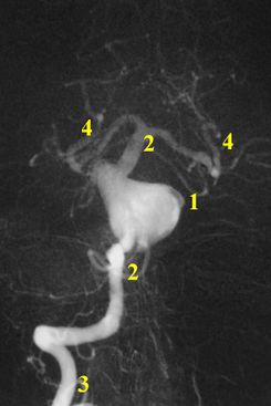
نزف تحت العنكبوتية Subarachnoid hemorrhage (SAH)، أو subarachnoid haemorrhage، هو نزيف داخل الحيز تحت العنكبوتية subarachnoid space المحيط بالمخ، وهي المنطقة بين الغشاء العنكبوني arachnoid membrane والأم الحنون pia mater. فيه يحدث النزف ضمن الطبقة السحائية العنكبوتية. وأكثر الأسباب شيوعاً هو رضوض الدماغ، أما حالات النزف غير الرَّضِّيّ فتنتج عادة من انفجار أم دم aneurysm (وهي توسع كيسي غير طبيعي في شريان أو أكثر). ومن الأسباب الأخرى التشوهات الشريانية الوريدية، والتهاب الأوعية، وتسلخ الشرايين، واعتلالات التخثر، وخثار الجيوب الوريدية، وفقر الدم المنجلي، وتمزق شريان سطحي صغير. ويبقى السبب مجهولاً في نحو 14ـ22% من الحالات. وفيما يأتي نبذة عن النزف تحت العنكبوتية الناتج من أمهات الدم لكونه الأهم بينها.[1]
. . . . . . . . . . . . . . . . . . . . . . . . . . . . . . . . . . . . . . . . . . . . . . . . . . . . . . . . . . . . . . . . . . . . . . . . . . . . . . . . . . . . . . . . . . . . . . . . . . . . . . . . . . . . . . . . . . . . . . . . . . . . . . . . . . . . . . . . . . . . . . . . . . . . . . . . . . . . . . . . . . . . . . . .
العلامات والأعراض
أهم الأعراض الصداع، وعادة ما يكون فجائياً وشديداً وكثيراً ما يترافق بالغثيان والإقياء والانزعاج من الضياء، والآلام في العنق. ومن الأعراض الأخرى حدوث إصابات بالأعصاب القحفية (ولاسيما الأعصاب المحركة للعين) وآلام أسفل الظهر.
التشخيص
| ||||||
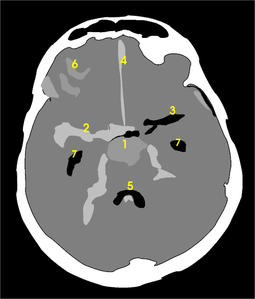
يعتمد بصفة رئيسة على تصوير الدماغ، ومن أكثر طرق التصوير استعمالاً التصوير الطبقي المحوري (الشكل3)، وبدرجة أقل المرنان المغنطيسي. وجدير بالذكر أن تصوير الدماغ قد يخفق في إظهار النزف في نحو 5% من المرضى، وفي هذه الحالة يمكن اللجوء إلى استقصاء آخر هو البزل القطني للسائل الدماغي الشوكي. وتظهر دراسة السائل وجود أعداد كبيرة من الكريات الحمر، مما يثبت حدوث النزف.
المسببات
Spontaneous SAH is most often due to rupture of cerebral aneurysms (85%), or weaknesses in the wall of the arteries of the brain that become enlarged. In 15-20% of cases of spontaneous SAH, no aneurysm is detected from the first angiogram.[2]
التصنيف
- مقياس هنت و هـِس Hunt and Hess scale
The first scale of severity, described by Hunt and Hess in 1968:[3]
- Grade 1: Asymptomatic; or minimal headache and slight nuchal rigidity. Approximate survival rate 70%.
- Grade 2: Moderate to severe headache; nuchal rigidity; no neurologic deficit except cranial nerve palsy. 60%.
- Grade 3: Drowsy; minimal neurologic deficit. 50%.
- Grade 4: Stuporous; moderate to severe hemiparesis; possibly early decerebrate rigidity and vegetative disturbances. 20%.
- Grade 5: Deep coma; decerebrate rigidity; moribund. 10%.
- درجات فيشر Fisher grade
The Fisher Grade classifies the appearance of subarachnoid hemorrhage on CT scan:[4]
- Grade 1: No hemorrhage evident
- Grade 2: Subarachnoid hemorrhage less than 1 mm thick
- Grade 3: Subarachnoid hemorrhage more than 1 mm thick
- Grade 4: Subarachnoid hemorrhage of any thickness with intra-ventricular hemorrhage (IVH) or parenchymal extension
- الاتحاد العالمي للجراحين العصبيين World Federation of Neurosurgeons
The World Federation of Neurosurgeons classification:[5]
- Class 1: GCS (Glasgow Coma Scale) 15
- Class 2: GCS 13-14 without focal neurological deficit
- Class 3: GCS 13-14 with focal neurological deficit
- Class 4: GCS 7-12 with or without focal neurological deficit
- Class 5: GCS <7 with or without focal neurological deficit
العلاج
الإجراءات العامة
The first priority is stabilization of the patient. In those with a depressed level of consciousness, intubation and mechanical ventilation may be required. Blood pressure, pulse, respiratory rate and Glasgow Coma Scale are monitored frequently. Once the diagnosis is confirmed, admission to an intensive care unit (ICU) may be considered preferable, especially given that 15% have a further episode (rebleeding) in the first hours after admission. Nutrition is an early priority, with oral or nasogastric tube feeding being preferable over parenteral routes. Analgesia (pain control) is generally restricted to non-sedating agents, as sedation would interfere with the monitoring of the level of consciousness. There is emphasis on the prevention of complications; for instance, deep vein thrombosis is prevented with compression stockings, intermittent pneumatic compression, or both.[6]
يعدّ النزف تحت العنكبوتية حالة إسعافية، ويؤدي إلى الوفاة في نحو نصف المرضى (وذلك في الحالات الناتجة من أم دم).
يتم عادة قبول المرضى في وحدة العناية المشددة لمراقبة ضغط الدم، وإعطاء السوائل الوريدية والأدوية المسكنة والمضادة للاختلاجات والمضادة للوذمة الدماغية (حسب الحالة)، وكذلك الأدوية المضادة للتشنج الشرياني (فقد يؤدي النزف إلى حدوث تخريش وتضيق في شرايين الدماغ مما قد يسبب حدوث نقص تروية دماغية)، أما العلاج الأساسي لمنع تكرار النزف فهو معالجة التشوه الوعائي في حال وجوده.
منع عودة النزف

المآل
SAH is often associated with a poor outcome.[7] The mortality rate for SAH is between 40 and 50%.[8] Between 10 and 20% of SAH patients that do not die before reaching the hospital die in the early weeks in hospital from rebleeding.[بحاجة لمصدر] Delay in diagnosis of minor SAH without coma (or mistaking the sudden headache for migraine) contributes to this mortality. Patients who remain comatose or with persistent severe deficits have a poor prognosis.[بحاجة لمصدر]
الوبائيات
Women have SAH more commonly than men do.[8] The group of people at risk for SAH is younger than the population usually affected by stroke.[7]
. . . . . . . . . . . . . . . . . . . . . . . . . . . . . . . . . . . . . . . . . . . . . . . . . . . . . . . . . . . . . . . . . . . . . . . . . . . . . . . . . . . . . . . . . . . . . . . . . . . . . . . . . . . . . . . . . . . . . . . . . . . . . . . . . . . . . . . . . . . . . . . . . . . . . . . . . . . . . . . . . . . . . . . .
انظر أيضاً
المصادر
- ^ محمد قتيبة الصفدي. "النزف السحائي". الموسوعة العربية. Retrieved 2011-05-23.
- ^ Rinkel GJ, van Gijn J, Wijdicks EF (1993). "Subarachnoid hemorrhage without detectable aneurysm. A review of the causes". Stroke. 24 (9): 1403–9. PMID 8362440.
{{cite journal}}: CS1 maint: multiple names: authors list (link) - ^ Hunt W, Hess R (1968). "Surgical risk as related to time of intervention in the repair of intracranial aneurysms". Journal of Neurosurgery. 28 (1): 14–20. PMID 5635959.
- ^ Fisher C, Kistler J, Davis J (1980). "Relation of cerebral vasospasm to subarachnoid hemorrhage visualized by computerized tomographic scanning". Neurosurgery. 6 (1): 1–9. PMID 7354892.
{{cite journal}}: CS1 maint: multiple names: authors list (link) - ^ Teasdale G, Drake C, Hunt W, Kassell N, Sano K, Pertuiset B, De Villiers J (1988). "A universal subarachnoid hemorrhage scale: Report of a committee of the World Federation of Neurosurgical Societies". J Neurol Neurosurg Psychiatry. 51 (11): 1457. PMID 3236024.
{{cite journal}}: CS1 maint: multiple names: authors list (link) - ^ خطأ استشهاد: وسم
<ref>غير صحيح؛ لا نص تم توفيره للمراجع المسماةvanGijn - ^ أ ب خطأ استشهاد: وسم
<ref>غير صحيح؛ لا نص تم توفيره للمراجع المسماةFeigin05 - ^ أ ب خطأ استشهاد: وسم
<ref>غير صحيح؛ لا نص تم توفيره للمراجع المسماةTeunissen96
وصلات خارجية
- Neuroland SAH page


