مجهر إلكتروني ماسح
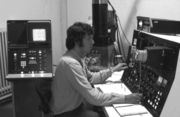
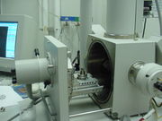
المجهر الإلكتروني الماسح ( Scanning Electron Microscope واختصاره SEM) هو نوع من المجاهر الإلكترونية التي تنتج صوراً بمسح السطح بشعاع مركز من الإلكترونات. تتفاعل الإلكترونات مع الذرة في العينة، وتنتج إشارات مختلفة تحتوي على معلومات عن طبوغرافيا السطح وتكوين العينة. يتم مسح شعاع الإلكترون في نمط المسح النقطي، ويتم دمج موضع الحزمة مع شدة الإشارة المكتشفة لإنتاج صورة. في وضع SEM الأكثر شيوعاً، يتم الكشف عن الإلكترونات الثانوية المنبعثة من الذرات التي تثيرها حزمة الإلكترون باستخدام كاشف إلكترون ثانوي (كاشف إڤرهارت-ثورنلي). يعتمد عدد الإلكترونات الثانوية التي يمكن الكشف عنها، وبالتالي شدة الإشارة، من بين أمور أخرى، على تضاريس العينة. يمكن لبعض SEMs تحقيق دقة أفضل من 1 نانومتر.
تـُرصـَد العينات في فراغ عالي في SEM تقليدي، أو في فراغ منخفض أو ظروف رطبة في ضغط متغير في SEM بيئي، وفي نطاق واسع من درجات الحرارة المبردة أو المرفوعة باستخدام أجهزة متخصصة.[1]
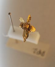
صمم هذا الميكروسكوب لفحص طوبوغرافية سطح العينة ، وهو يوفر قوة تكبير وقوة تحليل أقل مما يوفره الميكروسكوب الإلكتروني النافذ، ويستخدم على سبيل المثال لفحص أعين الحشرات وخياشيم الأسماك ..إلخ.
. . . . . . . . . . . . . . . . . . . . . . . . . . . . . . . . . . . . . . . . . . . . . . . . . . . . . . . . . . . . . . . . . . . . . . . . . . . . . . . . . . . . . . . . . . . . . . . . . . . . . . . . . . . . . . . . . . . . . . . . . . . . . . . . . . . . . . . . . . . . . . . . . . . . . . . . . . . . . . . . . . . . . . . .
عملية المسح

معرض صور SEM
The following are examples of images taken using a scanning electron microscope.
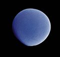
SEM Picture of a Diatom at magnification of 5000X.
False coloured SEM image of soybean cyst nematode and egg.
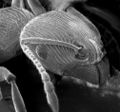
SEM image of an ant head.
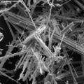
SEM image of asbestos fibers.
Compound eye of Antarctic krill Euphausia superba.
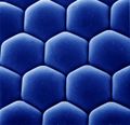
Ommatidia of Antarctic krill eye.
SEM image of a discharged nematocyst.
SEM image of the upper body of a Drosophila.
SEM images of the compound eye of a Drosophila.
SEM image of the grasshopper's spiracle valve.
SEM image of normal circulating human blood.
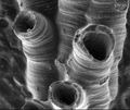
SEM image of a hederelloid from the Devonian of Michigan (largest tube diameter is 0.75 mm).
انظر أيضاً
المراجع
- ^ Stokes, Debbie J. (2008). Principles and Practice of Variable Pressure Environmental Scanning Electron Microscopy (VP-ESEM). Chichester: John Wiley & Sons. ISBN 978-0470758748.
وصلات خارجية
| مراجع مكتبية عن Scanning electron microscopy |
- عموميات
- HowStuffWorks – How Scanning Electron Microscopes Work
- Notes on the SEM Notes covering all aspects of the SEM
- Scanning Electron Microscopy basics an animated tutorial on how SEM works
- Learn to use an SEM – An online learning environment for people wanting to use an SEM. Provided by Microscopy Australia
- Virtual SEM – sparkler – an interactive simulation of a scanning electron microscope (SEM)
- Preparing a Sample for the SEM preparing a non-conducting subject for the SEM (QuickTime-movie)
- multichannel color SEM imaging – and with BSE
- DDC-SEM image examples
- Video on the scanning electron microscope, Karlsruhe University of Applied Sciences
- animations and explanations on various types of microscopes including electron microscopes (Université Paris Sud)
- تاريخ
- Microscopy History links from the University of Alabama Department of Biological Sciences
- Environmental Scanning Electron Microscope (ESEM) history
- صور
- Rippel Electron Microscope Facility Many dozens of (mostly biological) SEM images from Dartmouth College.
- Lanthanoid staining SEM images from Research Institute of Eye Diseases, Moscow.
