سيال عصبي

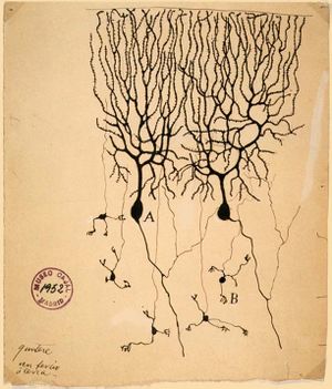
السيال العصبي أو النبض العصبي (Nerve Impulse) ، هو عملية نقل المعلومات أو النبضات العصبية داخل الأعصاب. وتتم عملية النقل إما بواسطة كهربائية (أنظر كمون الفعل) أو عن طريق التفاعلات الكيماوية بين الأعصاب. وإن سرعة السيال العصبي في الأعصاب تقدر بـ 120 متراً بالثانية أي ما يعادل 432 كم في الساعة.
يتكون الجهاز العصبي من خلايا منفردة، تتعاون معاً لإنجاز وظائف معقدة، وتدعى هذه الخلايا العصبونات. ان السيال العصبي هو اللغة الوحيدة التي تتفاهم بها العصبونات والشكل الذي تترجم إليه أنواع المؤثرات جميعها التي تؤثر في الجسم. ينتقل السيال من خلية عصبية لاخرى من خلال الروابط الفسيحة (gab junction) والتي هي عبارة عن قنوات دقيقة تسمح بسريان التيار من خلالها مباشرة وتكون هذه الطريقة أسرع مقارنة بالانتقال عبر التشابك الكيمياوي.
السياق الطبيعي الحيوي والخلوي
الأيونات والقوى التي تقود حركتهم
 مقالات مفصلة: أيون
- Diffusion
- Electrochemical gradient
- Electrophoretic mobility


غشاء الخلية

Membrane potential
قنوات الأيونات
مضخات الأيونات
Resting potential
حيث
- Eeq,K+ is the equilibrium potential for potassium, measured in volts
- R is the universal gas constant, equal to 8.314 joules·K-1·mol-1
- T is the absolute temperature, measured in kelvins (= K = degrees Celsius + 273.15)
- z is the number of elementary charges of the ion in question involved in the reaction
- F is the Faraday constant, equal to 96,485 coulombs·mol-1 or J·V-1·mol-1
- [K+]o is the extracellular concentration of potassium, measured in mol·m-3 or mmol·l-1
- [K+]i is the intracellular concentration of potassium

تشريح العصب
Initiation
Neurotransmission
الخلايا العصبية الحسية
Pacemaker potentials

الأطوار
Stimulation and rising phase
طور القمة والسقوط
Hyperpolarization ("undershoot")
Refractory period
الانتشار

Myelin and saltatory conduction

نظرية الكابل
 مقالة مفصلة: نظرية الكابل

where V(x, t) is the voltage across the membrane at a time t and a position x along the length of the neuron, and where λ and τ are the characteristic length and time scales on which those voltages decay in response to a stimulus. Referring to the circuit diagram above, these scales can be determined from the resistances and capacitances per unit length[6]
Termination
Chemical synapses

Electrical synapses
Neuromuscular junctions
أنواع أخرى من الخلايا
Cardiac action potentials

Muscular action potentials
Plant action potentials
التوزيع التصنيفي والمزايا التطورية
| Animal | Cell type | Resting potential (mV) | AP increase (mV) | AP duration (ms) | Conduction speed (m/s) |
|---|---|---|---|---|---|
| Squid (Loligo) | Giant axon | −60 | 120 | 0.75 | 35 |
| Earthworm (Lumbricus) | Median giant fiber | −70 | 100 | 1.0 | 30 |
| Cockroach (Periplaneta) | Giant fiber | −70 | 80–104 | 0.4 | 10 |
| Frog (Rana) | Sciatic nerve axon | −60 to −80 | 110–130 | 1.0 | 7–30 |
| Cat (Felis) | Spinal motor neuron | −55 to −80 | 80–110 | 1–1.5 | 30–120 |
طرق تجريبية


الذيفانات العصبية

التاريخ


نماذج كمية

انظر أيضا
- كمون فعل قلبي Cardiac action potential
- جهد غشائي Membrane potential
- دور الحران Refractory period
- زوال الاستقطاب Depolarization
- فرط الاستقطاب Hyperpolarization
- إشارة (أحياء)
- ثابت زمني Time constant
- ثابت الطول Length constant
- Bursting
- إشارات (علم الأحياء)
- Central pattern generator
ملاحظات
المصادر
- ^ Campbell Biology, 6th edition
- ^ CRC Handbook of Chemistry and Physics, 83rd edition, ISBN 0-8493-0483-0, pp. 12–14 to 12–16.
- ^ Schmidt-Nielsen, Figure 12.13.
- ^ خطأ استشهاد: وسم
<ref>غير صحيح؛ لا نص تم توفيره للمراجع المسماةhursh_1939 - ^ Rushton WAH (1951). "A theory of the effects of fibre size in the medullated nerve". Journal of Physiology. 115: 101–22.
- ^ Purves et al., pp. 52–53.
- ^ Bullock TH, Horridge GA (1965). Structure and Function in the Nervous Systems of Invertebrates. San Francisco: W. H. Freeman.
ببليوگرافيا
- Aidley DJ, Stanfield PR (1996). Ion Channels: Molecules in Action. Cambridge: Cambridge University Press. ISBN 978-0521498821.
- Bear MF, Connors BW, Paradiso MA (2001). Neuroscience: Exploring the Brain. Baltimore: Lippincott. ISBN 0781739446.
{{cite book}}: CS1 maint: multiple names: authors list (link) - Bullock TH, Orkand R, Grinnell A (1977). Introduction to Nervous Systems. New York: W. H. Freeman. ISBN 0-7167-0030-1.
{{cite book}}: CS1 maint: multiple names: authors list (link) - Clay JR (2005). "Axonal excitability revisited". Prog Biophys Mol Biol. 88 (1): 59–90. doi:10.1016/j.pbiomolbio.2003.12.004. PMID 15561301.
{{cite journal}}: Unknown parameter|month=ignored (help) - Deutsch S, Micheli-Tzanakou E (1987). Neuroelectric Systems. New York: New York University Press. ISBN 0-8147-1782-9.
- Hille B (2001). Ion Channels of Excitable Membranes (3rd ed.). Sunderland, MA: Sinauer Associates. ISBN 978-0878933211.
- Hoppensteadt FC (1986). An Introduction to the Mathematics of Neurons. Cambridge: Cambridge University Press. ISBN 0-521-31574-3.
- Johnston D, Wu SM-S (1995). Foundations of Cellular Neurophysiology. Cambridge, MA: Bradford Book, The MIT Press. ISBN 0-262-10053-3.
- Junge D (1981). Nerve and Muscle Excitation (2nd ed.). Sunderland MA: Sinauer Associates. ISBN 0-87893-410-3.
- Kandel ER, Schwartz JH, Jessell TM (2000). Principles of Neural Science (4th ed.). New York: McGraw-Hill. ISBN 0-8385-7701-6.
{{cite book}}: CS1 maint: multiple names: authors list (link) - Keynes RD, Aidley DJ (1991). Nerve and Muscle (2nd ed.). Cambridge: Cambridge University Press. ISBN 0-521-41042-8.
- Miller C (1987). "How ion channel proteins work". In LK Kaczmarek, IB Levitan (ed.). Neuromodulation: The Biochemical Control of Neuronal Excitability. New York: Oxford University Press. pp. 39–63. ISBN 978-0195040975.
- Nelson DL, Cox MM (2008). Lehninger Principles of Biochemistry (5th ed.). New York: W. H. Freeman. ISBN 978-0-7167-7108-1.
- Purves D, Augustine GJ, Fitzpatrick D, Hall WC, Lamantia A-S, McNamara JO, Williams SM (2001). "Release of Transmitters from Synaptic Vesicles". Neuroscience (2nd ed.). Sunderland, MA: Sinauer Associates. ISBN 0878937250.
{{cite book}}: External link in|chapterurl=|chapterurl=ignored (|chapter-url=suggested) (help)CS1 maint: multiple names: authors list (link) - Purves D, Augustine GJ, Fitzpatrick D, Hall WC, Lamantia A-S, McNamara JO, White LE (2008). Neuroscience (4th ed.). Sunderland, MA: Sinauer Associates. ISBN 978-0-87893-697-7.
{{cite book}}: CS1 maint: multiple names: authors list (link) - Schmidt-Nielsen K (1997). Animal Physiology: Adaptation and Environment (5th ed.). Cambridge: Cambridge University Press. ISBN 978-0521570985.
- Stevens CF (1966). Neurophysiology: A Primer. New York: John Wiley and Sons.LCCN 66-15872.
وصلات خارجية
- صور متحركة
- Ionic flow in action potentials at Blackwell Publishing
- Action potential propagation in myelinated and unmyelinated axons at Blackwell Publishing
- Generation of AP in cardiac cells and generation of AP in neuron cells
- Resting membrane potential from Life: The Science of Biology, by WK Purves, D Sadava, GH Orians, and HC Heller, 8th edition, New York: WH Freeman, ISBN 978-0716776710.
- Ionic motion and the Goldman voltage for arbitrary ionic concentrations at The University of Arizona
- A cartoon illustrating the action potential
- Action potential Propagation
- ملاحظات محاضرات ومواد أخرى
- The Action Potential John Kinnamon, University of Denver
- Resting and Action Membrane Potentials Teaching Resources Center, UC Davis. Animated tutorials
- Open-source software to simulate neuronal and cardiac action potentials at SourceForge.net
