ملف:Human skeletal muscle tissue 1 - TEM.jpg

الملف الأصلي (2٬200 × 1٬611 بكسل حجم الملف: 1٫01 ميجابايت، نوع MIME: image/jpeg)
وصف قصير
| ⧼wm-license-information-description⧽ |
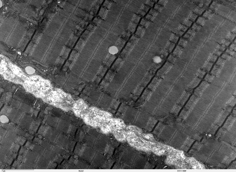
Transmission electron microscope image of a thin longitudinal section cut through an area of human skeletal muscle tissue. Image shows several myofibrils, each with the distinct banding pattern of individual sarcomeres. Image of muscle sarcomeres shows distinct banding pattern: the darker bands are called A bands(the A band includes a lighter central zone, called the H band), and the lighter bands are called I bands. Each I band is bisected by a dark transverse line called the Z-line). Paired mitochondria are on either side of the electron opaque Z-line. The Z-Line marks the longitudinal extent of a sarcomere unit. JEOL 100CX TEM |
| ⧼wm-license-information-date⧽ | |
| ⧼wm-license-information-source⧽ | |
| ⧼wm-license-information-author⧽ | Louisa Howard |
| ⧼wm-license-information-permission⧽ (⧼wm-license-information-permission-reusing-text⧽) |
PD |
ترخيص
|
|
This work has been released into the public domain by its author, Louisa Howard. This applies worldwide. In case this is not legally possible: |
تاريخ الملف
اضغط على زمن/تاريخ لرؤية الملف كما بدا في هذا الزمن.
| زمن/تاريخ | صورة مصغرة | الأبعاد | مستخدم | تعليق | |
|---|---|---|---|---|---|
| حالي | ★ مراجعة معتمدة 01:02، 12 نوفمبر 2023 | | 2٬200 × 1٬611 (1٫01 ميجابايت) | Pastakhov (نقاش | مساهمات) | Upload https://upload.wikimedia.org/wikipedia/commons/2/25/Human_skeletal_muscle_tissue_1_-_TEM.jpg |
لا يمكنك استبدال هذا الملف.
وصلات
لا يوجد صفحات تصل لهذه الصورة.