ملف:Human bocavirus 1 pneumonia.jpg
لا توجد دقة أعلى متوفرة.
Human_bocavirus_1_pneumonia.jpg (600 × 561 بكسل حجم الملف: 43 كيلوبايت، نوع MIME: image/jpeg)
| ⧼wm-license-information-description⧽ |
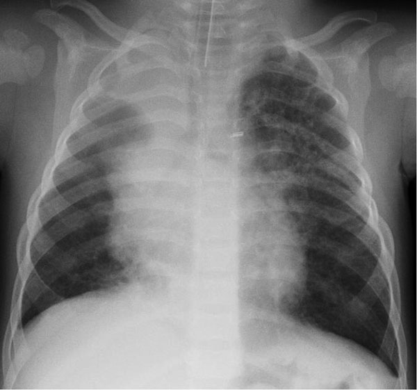
English: Figure. . Chest radiograph of the index patient, a 16-month-old boy in Finland with human bocavirus 1 pneumonia, on day 2 of hospitalization. Bilateral pulmonary infiltrations and atelectasis of the upper right lobe can be seen.
|
| ⧼wm-license-information-date⧽ | 2013 |
| ⧼wm-license-information-source⧽ | Emerging Infectious Diseases, Volume 19, Number 8—August 2013, http://wwwnc.cdc.gov/eid/article/19/8/13-0074-f1.htm |
| ⧼wm-license-information-author⧽ | Alma Jula, Matti Waris, Kalle Kantola, Ville Peltola, Maria Söderlund-Venermo, Klaus Hedman, and Olli Ruuskanen |
| ⧼wm-license-information-permission⧽ (⧼wm-license-information-permission-reusing-text⧽) |
تاريخ الملف
اضغط على زمن/تاريخ لرؤية الملف كما بدا في هذا الزمن.
| زمن/تاريخ | صورة مصغرة | الأبعاد | مستخدم | تعليق | |
|---|---|---|---|---|---|
| حالي | ★ مراجعة معتمدة 20:00، 14 نوفمبر 2023 | | 600 × 561 (43 كيلوبايت) | Pastakhov (نقاش | مساهمات) | Upload https://upload.wikimedia.org/wikipedia/commons/8/8a/Human_bocavirus_1_pneumonia.jpg |
لا يمكنك استبدال هذا الملف.
وصلات
لا يوجد صفحات تصل لهذه الصورة.