ملف:Histology bse.jpg
لا توجد دقة أعلى متوفرة.
Histology_bse.jpg (700 × 558 بكسل حجم الملف: 73 كيلوبايت، نوع MIME: image/jpeg)
| ⧼wm-license-information-description⧽ |
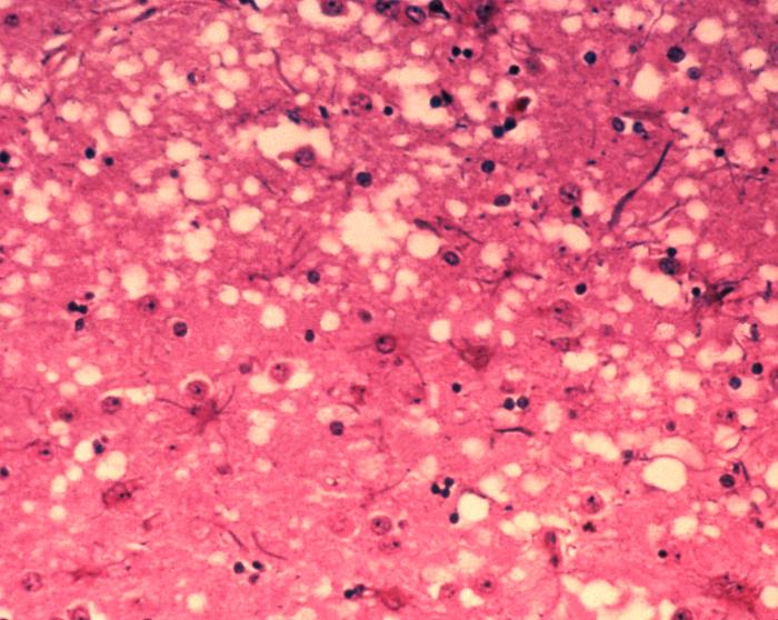
English: This micrograph of brain tissue reveals the cytoarchitectural histopathologic changes found in bovine spongiform encephalopathy. The presence of vacuoles, i.e. microscopic “holes” in the gray matter, gives the brain of BSE-affected cows a sponge-like appearance when tissue sections are examined in the lab.
(Dutch) Deutsch: Das Bild zeigt die histopathologischen Veränderungen die bei einer Infektion mit BSE auftreten. Die Vakuolen, die in der grauen Substanz (substantia grisea) auftreten geben dem Bild ein schwamm-artiges Aussehen.
Français : Cette coupe de tissu cérébral montre les modifications histopathologiques de l'organisation cellulaire lors d'une encéphalopathie spongiforme bovine. la présence de vacuoles, c'est-à-dire des "trous" microscopiques dans le tissu cérébral, donne au cerveau de vaches atteintes de l'ESB un aspect en éponge à l'examen des tissus en laboratoire.
|
| ⧼wm-license-information-date⧽ | 2003 |
| ⧼wm-license-information-source⧽ | Public Health Image Library, APHIS: http://www.aphis.usda.gov/lpa/issues/bse/bse_photogallery.html |
| ⧼wm-license-information-author⧽ | Dr. Al Jenny |
| ⧼wm-license-information-other-versions⧽ |
تاريخ الملف
اضغط على زمن/تاريخ لرؤية الملف كما بدا في هذا الزمن.
| زمن/تاريخ | صورة مصغرة | الأبعاد | مستخدم | تعليق | |
|---|---|---|---|---|---|
| حالي | ★ مراجعة معتمدة 11:43، 30 نوفمبر 2023 | | 700 × 558 (73 كيلوبايت) | Pastakhov (نقاش | مساهمات) | Upload https://upload.wikimedia.org/wikipedia/commons/2/23/Histology_bse.jpg |
لا يمكنك استبدال هذا الملف.
وصلات
لا يوجد صفحات تصل لهذه الصورة.