ملف:Hamartoma of the spleen.jpg
Hamartoma_of_the_spleen.jpg (537 × 376 بكسل حجم الملف: 50 كيلوبايت، نوع MIME: image/jpeg)
وصف قصير
| ⧼wm-license-information-description⧽ |
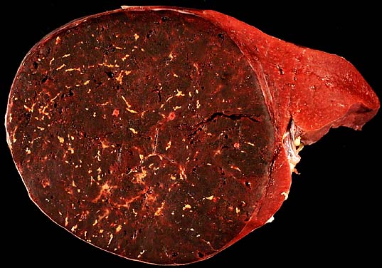
Hamartoma of the spleen A young man presented with left upper quadrant abdominal pain. On evaluation, a CT scan showed an enlarged spleen. He was worked up for lymphoma/leukemia, but nothing abnormal was found. CBC and bone marrow biopsy were normal. He was referred to a general surgeon for splenectomy. The specimen was a spleen containing a solitary 9-cm dark red mass that protruded above the surface of the adjacent spleen on section. The photo shows a slice through the specimen to show the tumor on the left and the normal splenic tissue on the right of the image. These fairly rare lesions are usually smaller and discovered incidentally at autopsy or splenectomy for unrelated purpose, but some larger lesions can produce pain. Unlike angiomas of the spleen, hamartomas rarely cause peripheral cytopenias. Photograph by Ed Uthman, MD. Public domain. Posted 2 Jan 99 |
| ⧼wm-license-information-date⧽ | |
| ⧼wm-license-information-source⧽ | http://web2.airmail.net/uthman/specimens/index.html |
| ⧼wm-license-information-author⧽ | |
| ⧼wm-license-information-permission⧽ (⧼wm-license-information-permission-reusing-text⧽) |
PD |
ترخيص
|
|
This work has been released into the public domain by its author, Ed Uthman. This applies worldwide. In case this is not legally possible: |
تاريخ الملف
اضغط على زمن/تاريخ لرؤية الملف كما بدا في هذا الزمن.
| زمن/تاريخ | صورة مصغرة | الأبعاد | مستخدم | تعليق | |
|---|---|---|---|---|---|
| حالي | ★ مراجعة معتمدة 11:01، 11 أكتوبر 2023 | | 537 × 376 (50 كيلوبايت) | Pastakhov (نقاش | مساهمات) | Upload https://upload.wikimedia.org/wikipedia/commons/7/7a/Hamartoma_of_the_spleen.jpg |
لا يمكنك استبدال هذا الملف.
وصلات
لا يوجد صفحات تصل لهذه الصورة.