ملف:Functional magnetic resonance imaging.jpg
لا توجد دقة أعلى متوفرة.
Functional_magnetic_resonance_imaging.jpg (250 × 208 بكسل حجم الملف: 11 كيلوبايت، نوع MIME: image/jpeg)
وصف قصير
| ⧼wm-license-information-description⧽ |
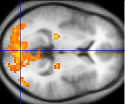
English: Sample fMRI data
This example of fMRI data shows regions of activation including primary visual cortex (V1, BA17), extrastriate visual cortex and lateral geniculate body in a comparison between a task involving a complex moving visual stimulus and rest condition (viewing a black screen). The activations (yellow-red) are shown (as is typical) against a background based on the average structural images from the subjects in the experiment. |
| ⧼wm-license-information-date⧽ | 2004 |
| ⧼wm-license-information-source⧽ |
|
| ⧼wm-license-information-author⧽ | قالب:User at project |
ترخيص
| [ This image has been (or is hereby) released into the public domain by its author, Washington irving at the English Wikipedia project. This applies worldwide. In case this is not legally possible: Deutsch · Ελληνικά · English · Plattdüütsch · 中文(简体) · 中文(繁體) · +/− |
قالب:Original upload log
قالب:Original file page
Public-domain: copyright disclaimed Washington irving 07:49, 8 Mar 2004 (UTC)
تاريخ الملف
اضغط على زمن/تاريخ لرؤية الملف كما بدا في هذا الزمن.
| زمن/تاريخ | صورة مصغرة | الأبعاد | مستخدم | تعليق | |
|---|---|---|---|---|---|
| حالي | ★ مراجعة معتمدة 22:21، 5 أكتوبر 2023 | | 250 × 208 (11 كيلوبايت) | Pastakhov (نقاش | مساهمات) | Upload https://upload.wikimedia.org/wikipedia/commons/8/87/Functional_magnetic_resonance_imaging.jpg |
لا يمكنك استبدال هذا الملف.
وصلات
لا يوجد صفحات تصل لهذه الصورة.