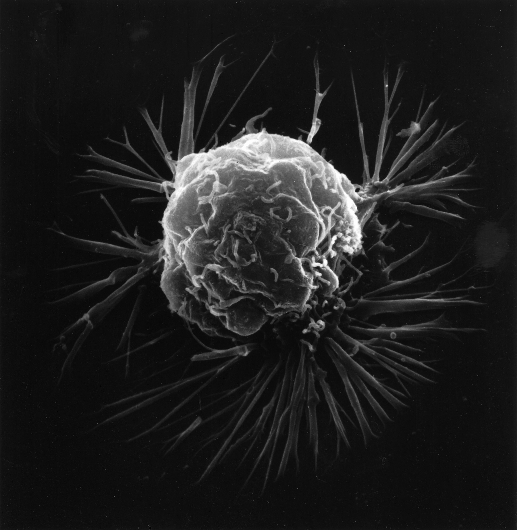
ملف:Breast cancer cell (2).jpg
حجم هذه المعاينة: 585 × 599 بكسل. البعد الآخر: 1٬800 × 1٬844 بكسل.
الملف الأصلي (1٬800 × 1٬844 بكسل حجم الملف: 642 كيلوبايت، نوع MIME: image/jpeg)
وصف قصير
| ⧼wm-license-information-description⧽ |
English:
|
| ⧼wm-license-information-date⧽ | 1980 |
| ⧼wm-license-information-source⧽ | قالب:NCI Visuals Online |
| ⧼wm-license-information-author⧽ | قالب:Unknown photographer |
| ⧼wm-license-information-permission⧽ (⧼wm-license-information-permission-reusing-text⧽) |
Reuse Restrictions None - This image is in the public domain and can be freely reused. Please credit the source and/or author listed above. |
ترخيص
تاريخ الملف
اضغط على زمن/تاريخ لرؤية الملف كما بدا في هذا الزمن.
| زمن/تاريخ | صورة مصغرة | الأبعاد | مستخدم | تعليق | |
|---|---|---|---|---|---|
| حالي | ★ مراجعة معتمدة 04:56، 9 أكتوبر 2023 | | 1٬800 × 1٬844 (642 كيلوبايت) | Pastakhov (نقاش | مساهمات) | Upload https://upload.wikimedia.org/wikipedia/commons/b/bd/Breast_cancer_cell_%282%29.jpg |
لا يمكنك استبدال هذا الملف.
وصلات
لا يوجد صفحات تصل لهذه الصورة.
.jpg&oldid=3032626)