ملف:Appendicular skeleton diagram.svg

حجم معاينة PNG لذلك الملف ذي الامتداد SVG: 324 × 599 بكسل. البعد الآخر: 1٬107 × 2٬048 بكسل.
الملف الأصلي (ملف SVG، أبعاده 437 × 808 بكسل، حجم الملف: 1٫36 ميجابايت)
| ⧼wm-license-information-description⧽ |
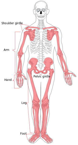
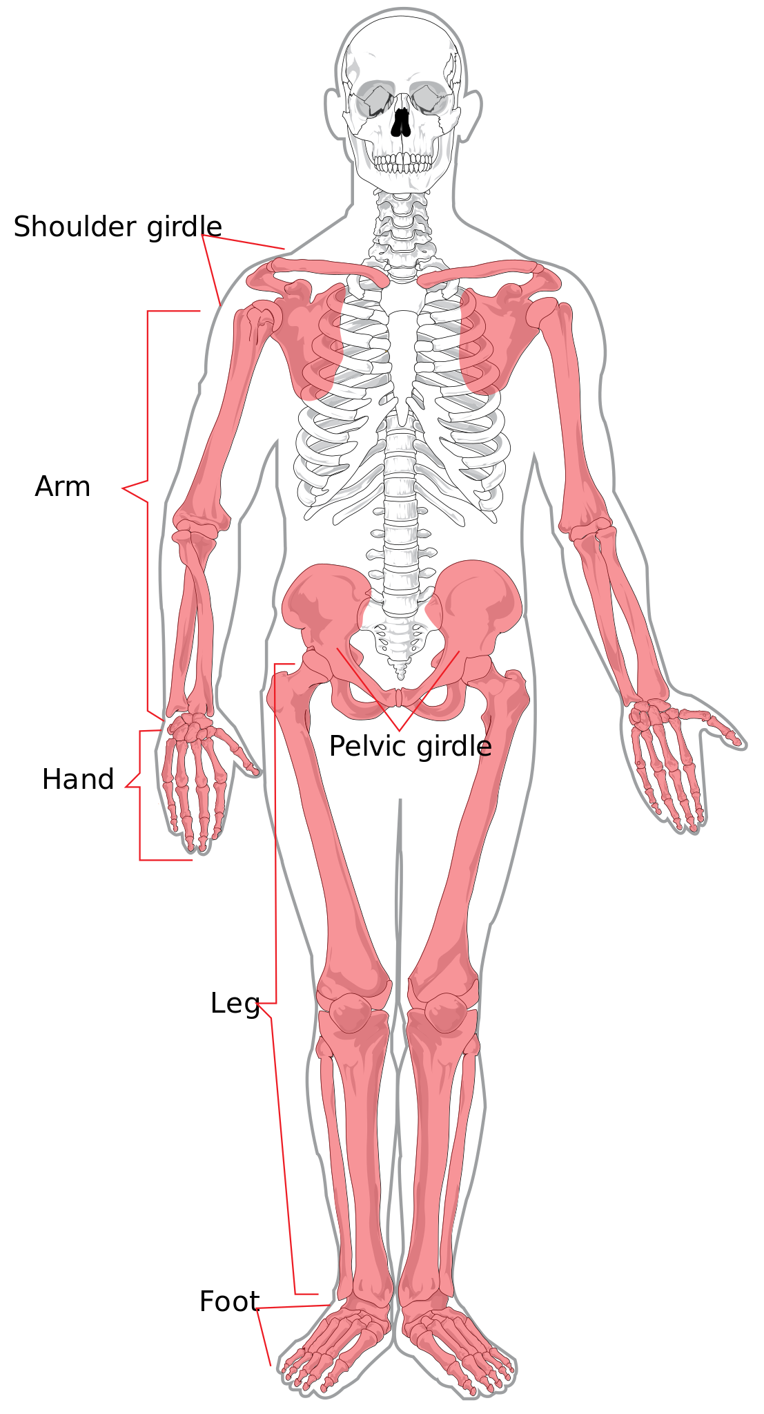
The appendicular skeleton and the axial skeleton together form the complete skeleton. Please note this diagram omits the intermediate phalanges of the 2nd-5th toes on both feet (the great toe does not have an intermediate phalanx). |
| ⧼wm-license-information-date⧽ | 2007 |
| ⧼wm-license-information-source⧽ | i did it myself |
| ⧼wm-license-information-author⧽ | LadyofHats Mariana Ruiz Villarreal |
| ⧼wm-license-information-permission⧽ (⧼wm-license-information-permission-reusing-text⧽) |
|
| ⧼wm-license-information-other-versions⧽ |
تاريخ الملف
اضغط على زمن/تاريخ لرؤية الملف كما بدا في هذا الزمن.
| زمن/تاريخ | صورة مصغرة | الأبعاد | مستخدم | تعليق | |
|---|---|---|---|---|---|
| حالي | ★ مراجعة معتمدة 23:03، 10 أكتوبر 2023 | | 437 × 808 (1٫36 ميجابايت) | Pastakhov (نقاش | مساهمات) | Upload https://upload.wikimedia.org/wikipedia/commons/7/7c/Appendicular_skeleton_diagram.svg |
لا يمكنك استبدال هذا الملف.
وصلات
لا يوجد صفحات تصل لهذه الصورة.