اللولب المزدوج للحمض النووي
في علم الأحياء الجزيئي، يشير مصطلح اللولب المزدوج double helix [1] إلى البنية المشكلة بواسطة الجزيئيات المزدوجة للأحماض النووية مثل الدنا. تنشأ البنية اللولبية المزدوجة لمركب الحمض النووي نتيجة لبنيته الثانوية، وتعتبر مكوناً أساسياً في تحديد لبنيته الثالثية. دخل المصطلح الثقافة العامة عام 1968 مع نشر اللولب المزدوج: سرد شخصي لاكتشاف بنية الدنا، د. جيمس واتسون.
يتجمع پوليمر اللولب المزدوج لدنا الحمض النووي عن طريق النيوكليوتايدات وتشكلان زوجاً قاعدياً.[2] B-DNA هي البنية اللولبية المزدوجة الأكثر شيوعاً في الطبيعة، اللولب المزدوج is right-handed بحوالي 10-10.5 زوج قاعدي لكل لفة.[3] تحتوي بنية اللولب المزدوج للدنا على أخدود رئيسي وأخدود أصغر. في B-DNA يكون الأخدود الرئيسي أعرض من الأخدود الأصغر.[2] نظرا للاختلاف في عرض الأخدود الرئيسي والأخدود الصغير، العديد من الپروتينات التي ترتبط ببنية B-DNA تفعل ذلك من خلال الأخدود الرئيسي الأعرض.[4]
التاريخ
نُشر نموجذ اللولب المزدوج للدنا أول مرة في مجلة نيتشر بواسطة جيمس واتسون وفرانسيس كريك عام 1953،[5] (الاحداثيات X، Y، Z عام 1954[6]) استنادا إلى صورة حيود الأشعة السينية حاسمة للدنا تحمل اسم "الصورة 51" من روزالند فرانكلن عام 1952،[7] تلتها صورة أكثر توضيحاً للدنا صنعتها فرانكلن ريموند گوسلنگ،[8][9] موريس ويلكينز، ألكس ستوكس، وهربرت ويلسون،[10] والمعلومات الكيميائية والكيميائية الحيوية للاقتران القاعدي من إروين شارگاف.[11][12][13][14][15][16] النموذج السابق كان triple-stranded DNA.[17]
تهجين الحمض النووي
هندسة الزوج القاعدي
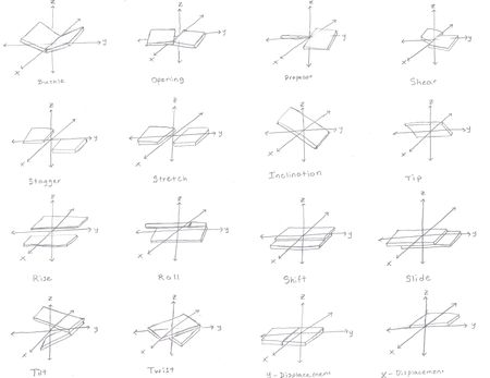
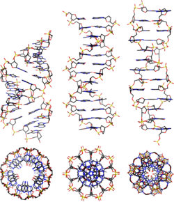
هندسة اللولب
| Geometry attribute | A-DNA | B-DNA | Z-DNA |
|---|---|---|---|
| Helix sense | right-handed | right-handed | left-handed |
| Repeating unit | 1 bp | 1 bp | 2 bp |
| Rotation/bp | 32.7° | 34.3° | 60°/2 |
| bp/turn | 11 | 10.5 | 12 |
| Inclination of bp to axis | +19° | −1.2° | −9° |
| Rise/bp along axis | 2.3 Å (0.23 nm) | 3.32 Å (0.332 nm) | 3.8 Å (0.38 nm) |
| Pitch/turn of helix | 28.2 Å (2.82 nm) | 33.2 Å (3.32 nm) | 45.6 Å (4.56 nm) |
| Mean propeller twist | +18° | +16° | 0° |
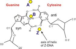
| Glycosyl angle | anti | anti | C: anti, G: syn |
| Sugar pucker | C3'-endo | C2'-endo | C: C2'-endo, G: C2'-exo |
| Diameter | 23 Å (2.3 nm) | 20 Å (2.0 nm) | 18 Å (1.8 nm) |
الأخاديد

الأشكال اللولبية الغير مزدوجة
الانحناء
الطول الثابت والصلابة المحورية
| التسلسل | الطول الثابت / الزوج القاعدي |
|---|---|
| العشوائية | 154±10 |
| (CA)التكرار | 133±10 |
| (CAG)التكرار | 124±10 |
| (TATA)التكرار | 137±10 |
أنماط تقوس الدنا
| Step | Stacking ΔG /kcal mol−1 |
|---|---|
| T A | -0.19 |
| T G or C A | -0.55 |
| C G | -0.91 |
| A G or C T | -1.06 |
| A A or T T | -1.11 |
| A T | -1.34 |
| G A or T C | -1.43 |
| C C or G G | -1.44 |
| A C or G T | -1.81 |
| G C | -2.17 |
أولوية الانحناء
| ¦ | ¦ | ¦ | ¦ | ¦ | ¦ | |||||||||||||||||||||||||||||||||||||||||||||||||||||||||||||||||||||||||||||||||||||||||||||||
| G | A | T | T | C | C | C | A | A | A | A | A | T | G | T | C | A | A | A | A | A | A | T | A | G | G | C | A | A | A | A | A | A | T | G | C | C | A | A | A | A | A | A | T | C | C | C | A | A | A | C |
التعميم
التمدد
نظام التمدد المرن
مرحلة انتقالات تحت التمدد
اللف الفائق والطبولوجيا
مفارقة رقم الارتباط
انظر أيضاً
- Triple-stranded DNA
- G-quadruplex
- التكنولوجيا النانوية للدنا
- النماذج الجزيئية للدنا
- البنية الجزيئية للحمض النووي (منشور)
- مقارنة برمجيات محاكاة الحمض النووي
المصادر
- ^ Kabai, Sándor (2007). "Double Helix". The Wolfram Demonstrations Project.
- ^ أ ب Alberts; et al. (1994). The Molecular Biology of the Cell. New York: Garland Science. ISBN 978-0-8153-4105-5.
- ^ Wang JC (1979). "Helical repeat of DNA in solution". PNAS. 76 (1): 200–203. Bibcode:1979PNAS...76..200W. doi:10.1073/pnas.76.1.200. PMC 382905. PMID 284332.
- ^ Pabo C, Sauer R (1984). "Protein-DNA recognition". Annu Rev Biochem. 53: 293–321. doi:10.1146/annurev.bi.53.070184.001453. PMID 6236744.
- ^ James Watson and Francis Crick (1953). "A structure for deoxyribose nucleic acid" (PDF). Nature. 171 (4356): 737–738. Bibcode:1953Natur.171..737W. doi:10.1038/171737a0. PMID 13054692.
- ^ Crick F, Watson JD (1954). "The Complementary Structure of Deoxyribonucleic Acid" (PDF). Proceedings of the Royal Society of London. 223, Series A: 80–96.
- ^ "Secret of Photo 51". Nova. PBS.
- ^ http://www.nature.com/nature/dna50/franklingosling.pdf
- ^ The Structure of the DNA Molecule Archived 2012-06-21 at the Wayback Machine
- ^ Wilkins MH, Stokes AR, Wilson HR (1953). "Molecular Structure of Deoxypentose Nucleic Acids" (PDF). Nature. 171 (4356): 738–740. Bibcode:1953Natur.171..738W. doi:10.1038/171738a0. PMID 13054693.
- ^ Elson D, Chargaff E (1952). "On the deoxyribonucleic acid content of sea urchin gametes". Experientia. 8 (4): 143–145. doi:10.1007/BF02170221. PMID 14945441.
- ^ Chargaff E, Lipshitz R, Green C (1952). "Composition of the deoxypentose nucleic acids of four genera of sea-urchin". J Biol Chem. 195 (1): 155–160. PMID 14938364.
- ^ Chargaff E, Lipshitz R, Green C, Hodes ME (1951). "The composition of the deoxyribonucleic acid of salmon sperm". J Biol Chem. 192 (1): 223–230. PMID 14917668.
- ^ Chargaff E (1951). "Some recent studies on the composition and structure of nucleic acids". J Cell Physiol Suppl. 38 (Suppl).
- ^ Magasanik B, Vischer E, Doniger R, Elson D, Chargaff E (1950). "The separation and estimation of ribonucleotides in minute quantities". J Biol Chem. 186 (1): 37–50. PMID 14778802.
- ^ Chargaff E (1950). "Chemical specificity of nucleic acids and mechanism of their enzymatic degradation". Experientia. 6 (6): 201–209. doi:10.1007/BF02173653. PMID 15421335.
- ^ Pauling L, Corey RB (Feb 1953). "A proposed structure for the nucleic acids". Proc Natl Acad Sci U S A. 39 (2): 84–97. Bibcode:1953PNAS...39...84P. doi:10.1073/pnas.39.2.84. PMC 1063734. PMID 16578429.
- ^ Rich A, Norheim A, Wang AH (1984). "The chemistry and biology of left-handed Z-DNA". Annual Review of Biochemistry. 53: 791–846. doi:10.1146/annurev.bi.53.070184.004043. PMID 6383204.
- ^ Sinden, Richard R (1994-01-15). DNA structure and function (1st ed.). Academic Press. p. 398. ISBN 0-12-645750-6.
- ^ Ho PS (1994-09-27). "The non-B-DNA structure of d(CA/TG)n does not differ from that of Z-DNA". Proc Natl Acad Sci USA. 91 (20): 9549–9553. Bibcode:1994PNAS...91.9549H. doi:10.1073/pnas.91.20.9549. PMC 44850. PMID 7937803.
- ^ Protozanova E, Yakovchuk P, Frank-Kamenetskii MD (2004). "Stacked–Unstacked Equilibrium at the Nick Site of DNA". J Mol Biol. 342 (3): 775–785. doi:10.1016/j.jmb.2004.07.075. PMID 15342236.


