ملف:Rhodopsin 3D.jpeg
حجم هذه المعاينة: 386 × 599 بكسل. البعد الآخر: 445 × 690 بكسل.
الملف الأصلي (445 × 690 بكسل حجم الملف: 49 كيلوبايت، نوع MIME: image/jpeg)
وصف قصير
| ⧼wm-license-information-description⧽ |
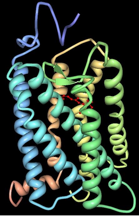
Structure cristallographique 3D de la Rhodopsine bovine (#1F88). |
| ⧼wm-license-information-date⧽ | 2008 |
| ⧼wm-license-information-source⧽ | Palczewski K, et al ("Crystal structure of rhodopsin: A G protein-coupled receptor." Science. 2000 Aug 4;289(5480):739-45
NOTE: Originally uploaded 06:06, 25 June 2006 (UTC) (log) to en:wikipedia by Roland Deschain (talk • contribs) with the following summary:
|
| ⧼wm-license-information-author⧽ | Roland Deschain |
ترخيص
تاريخ الملف
اضغط على زمن/تاريخ لرؤية الملف كما بدا في هذا الزمن.
| زمن/تاريخ | صورة مصغرة | الأبعاد | مستخدم | تعليق | |
|---|---|---|---|---|---|
| حالي | ★ مراجعة معتمدة 18:26، 15 أكتوبر 2023 | | 445 × 690 (49 كيلوبايت) | Pastakhov (نقاش | مساهمات) | Upload https://upload.wikimedia.org/wikipedia/commons/8/84/Rhodopsin_3D.jpeg |
لا يمكنك استبدال هذا الملف.
وصلات
لا يوجد صفحات تصل لهذه الصورة.