ملف:IL12 Crystal Structure.rsh.png
حجم هذه المعاينة: 800 × 589 بكسل. البعد الآخر: 1٬018 × 750 بكسل.
الملف الأصلي (1٬018 × 750 بكسل حجم الملف: 242 كيلوبايت، نوع MIME: image/png)
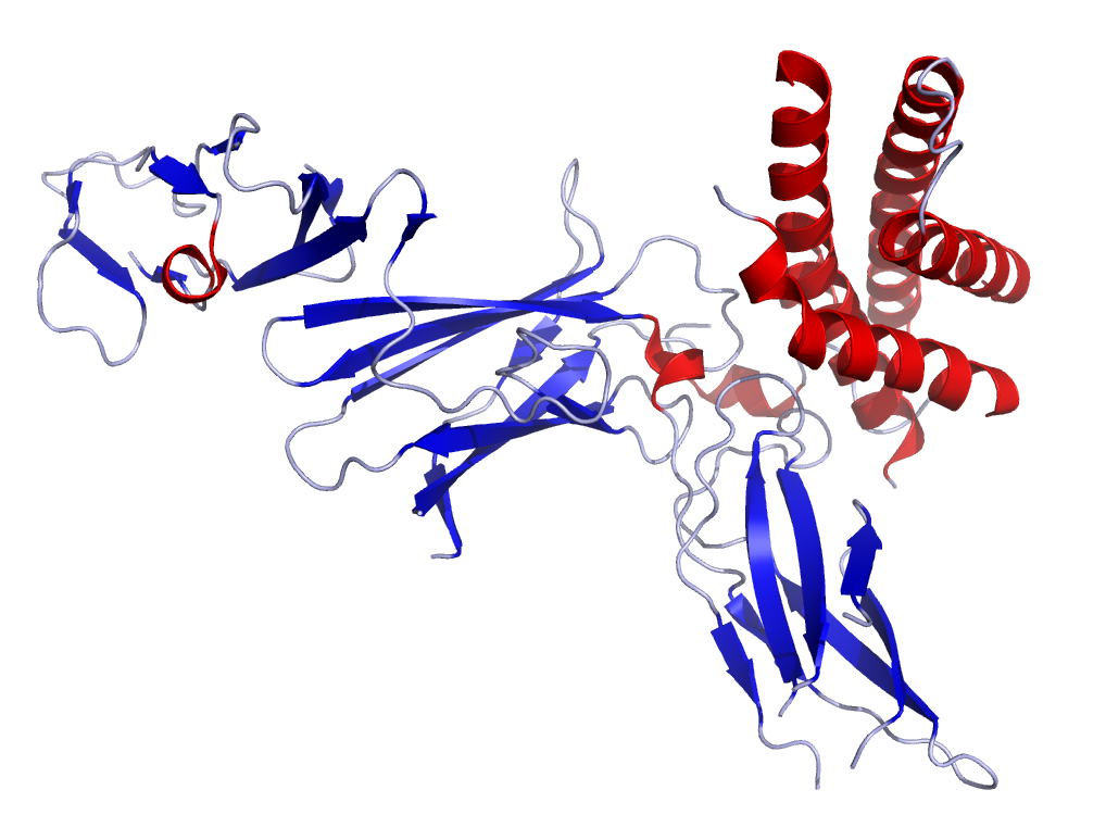
This is the crystal structure to human Interleukin-12. I accessed the data from the Protein Data Bank (PDB: 1F45) and rendered it using Pymol.
For the original data, see: Yoon, C., Johnston, S.C., Tang, J., Stahl, M., Tobin, J.F., Somers, W.S. Charged residues dominate a unique interlocking topography in the heterodimeric cytokine interleukin-12. EMBO J. v19 pp.3530-3541, 2000.
| ⧼wm-license-information-description⧽ |
Crystal structure of IL-12 as published in the Protein Data Bank (PDB: 1F45) |
||
| ⧼wm-license-information-date⧽ | 2006 | ||
| ⧼wm-license-information-source⧽ | Created from PDB 1F45 and rendered by me using Pymol | ||
| ⧼wm-license-information-author⧽ | Ramin Herati | ||
| ⧼wm-license-information-permission⧽ (⧼wm-license-information-permission-reusing-text⧽) |
|
تاريخ الملف
اضغط على زمن/تاريخ لرؤية الملف كما بدا في هذا الزمن.
| زمن/تاريخ | صورة مصغرة | الأبعاد | مستخدم | تعليق | |
|---|---|---|---|---|---|
| حالي | ★ مراجعة معتمدة 05:46، 19 أكتوبر 2023 | | 1٬018 × 750 (242 كيلوبايت) | Pastakhov (نقاش | مساهمات) | Upload https://upload.wikimedia.org/wikipedia/commons/5/55/IL12_Crystal_Structure.rsh.png |
لا يمكنك استبدال هذا الملف.
وصلات
لا يوجد صفحات تصل لهذه الصورة.