ملف:Herpes simplex cytopathy.jpg
حجم هذه المعاينة: 800 × 450 بكسل. البعد الآخر: 960 × 540 بكسل.
الملف الأصلي (960 × 540 بكسل حجم الملف: 69 كيلوبايت، نوع MIME: image/jpeg)
وصف قصير
| ⧼wm-license-information-description⧽ |
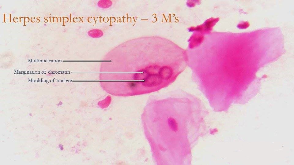
English: Pap smear -Multinucleated giant cell representing infection by Herpes simplex virus.The cell is multinucleated and the nuclei appear moulded (lost the normal round contour and flattened where they are in contact with each other,i.e moulded like clay balls pressed together).The virus replicates inside the nucleus and pushes the chromatin of the nucleus to its periphery(seen as a dark rim near nuclear membrane i.e Margination of chromatin).
|
| ⧼wm-license-information-date⧽ | 2017 |
| ⧼wm-license-information-source⧽ | Department of Pathology, Government Medical College, Kozikode |
| ⧼wm-license-information-author⧽ | Dr. Roshan Nasimudeen |
| ⧼wm-license-information-permission⧽ (⧼wm-license-information-permission-reusing-text⧽) |
ترخيص
|
تاريخ الملف
اضغط على زمن/تاريخ لرؤية الملف كما بدا في هذا الزمن.
| زمن/تاريخ | صورة مصغرة | الأبعاد | مستخدم | تعليق | |
|---|---|---|---|---|---|
| حالي | ★ مراجعة معتمدة 05:27، 11 أكتوبر 2023 | | 960 × 540 (69 كيلوبايت) | Pastakhov (نقاش | مساهمات) | Upload https://upload.wikimedia.org/wikipedia/commons/7/7b/Herpes_simplex_cytopathy.jpg |
لا يمكنك استبدال هذا الملف.
وصلات
لا يوجد صفحات تصل لهذه الصورة.