ملف:Chlamydomonas TEM 16.jpg
الملف الأصلي (1٬650 × 1٬318 بكسل حجم الملف: 732 كيلوبايت، نوع MIME: image/jpeg)
| ⧼wm-license-information-description⧽ |
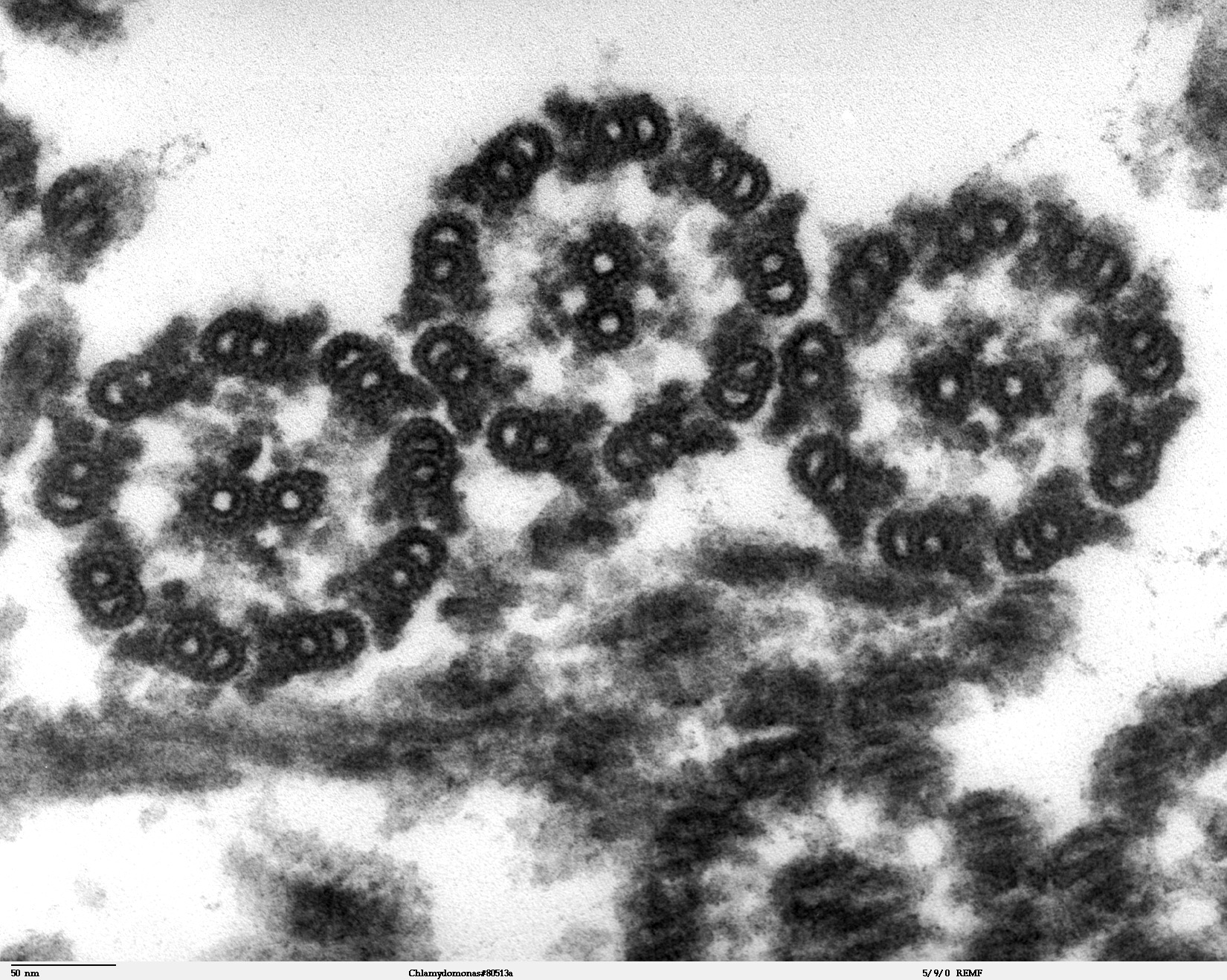
Transmission electron microscope image, showing an example of green algae (Chlorophyta). |
| ⧼wm-license-information-date⧽ | 2007 |
| ⧼wm-license-information-source⧽ | Source and public domain notice at http://remf.dartmouth.edu/imagesindex.html |
| ⧼wm-license-information-author⧽ | Dartmouth Electron Microscope Facility, Dartmouth College |
| ⧼wm-license-information-permission⧽ (⧼wm-license-information-permission-reusing-text⧽) |
Released into the public domain |
|
|
This work has been released into the public domain by its author, Dartmouth Electron Microscope Facility, Dartmouth College. This applies worldwide. In case this is not legally possible: |
تاريخ الملف
اضغط على زمن/تاريخ لرؤية الملف كما بدا في هذا الزمن.
| زمن/تاريخ | صورة مصغرة | الأبعاد | مستخدم | تعليق | |
|---|---|---|---|---|---|
| حالي | ★ مراجعة معتمدة 23:16، 30 نوفمبر 2023 | | 1٬650 × 1٬318 (732 كيلوبايت) | Pastakhov (نقاش | مساهمات) | Upload https://upload.wikimedia.org/wikipedia/commons/8/82/Chlamydomonas_TEM_16.jpg |
لا يمكنك استبدال هذا الملف.
وصلات
لا يوجد صفحات تصل لهذه الصورة.