ملف:Castleman disease - high mag.jpg
حجم هذه المعاينة: 400 × 600 بكسل. البعدان الآخران: 1٬365 × 2٬048 بكسل | 2٬848 × 4٬272 بكسل.
الملف الأصلي (2٬848 × 4٬272 بكسل حجم الملف: 6٫57 ميجابايت، نوع MIME: image/jpeg)
وصف قصير
| ⧼wm-license-information-description⧽ |
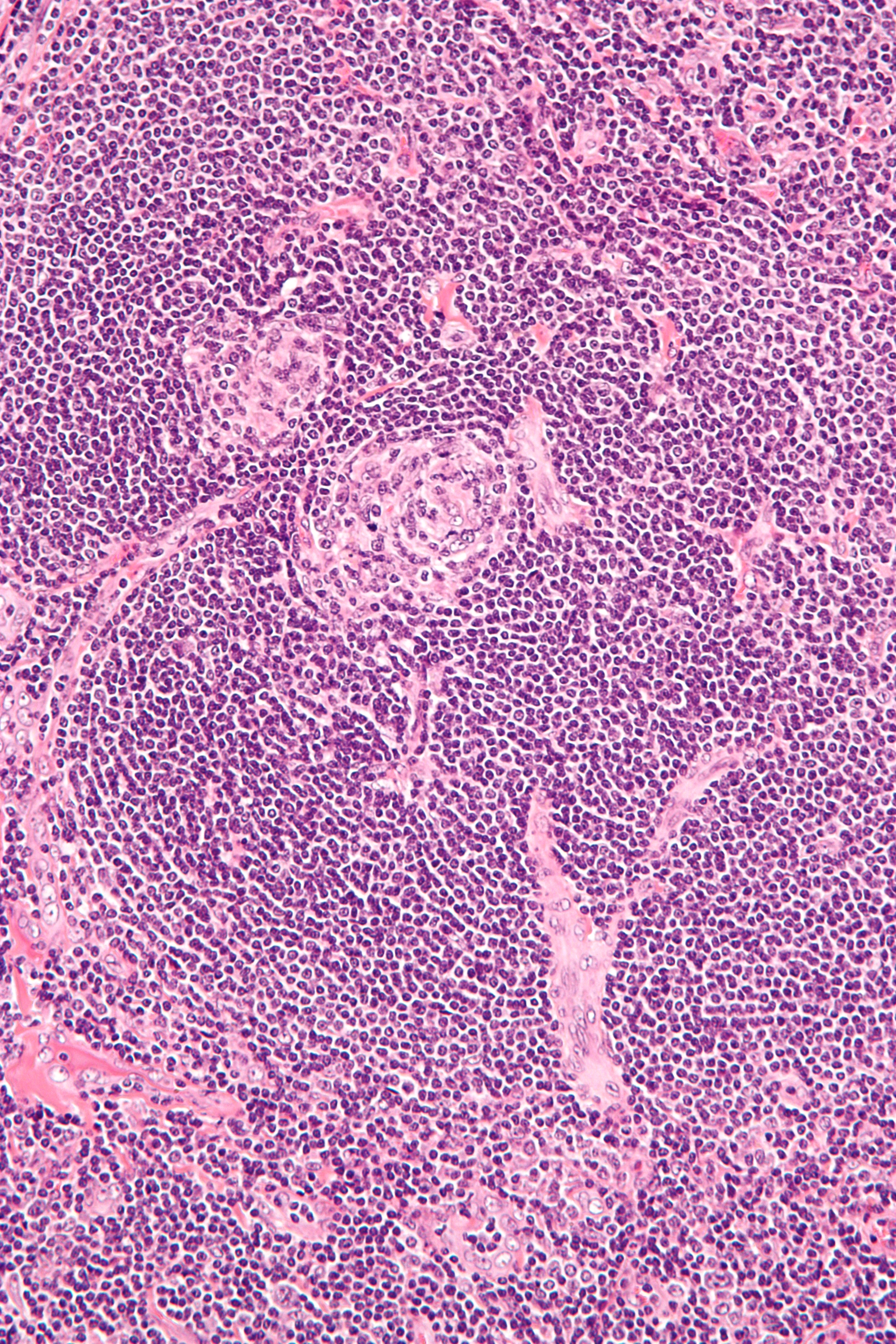
English: High magnification micrograph of Castleman disease, hyaline vascular variant, also known as angiofollicular lymph node hyperplasia and giant lymph node hyperplasia. H&E stain.
Features:
Related images
|
| ⧼wm-license-information-date⧽ | |
| ⧼wm-license-information-source⧽ | ⧼Wm-license-own-work⧽ |
| ⧼wm-license-information-author⧽ | Nephron |
ترخيص
|

تاريخ الملف
اضغط على زمن/تاريخ لرؤية الملف كما بدا في هذا الزمن.
| زمن/تاريخ | صورة مصغرة | الأبعاد | مستخدم | تعليق | |
|---|---|---|---|---|---|
| حالي | ★ مراجعة معتمدة 10:16، 2 نوفمبر 2023 | | 2٬848 × 4٬272 (6٫57 ميجابايت) | Pastakhov (نقاش | مساهمات) | Upload https://upload.wikimedia.org/wikipedia/commons/6/66/Castleman_disease_-_high_mag.jpg |
لا يمكنك استبدال هذا الملف.
وصلات
لا يوجد صفحات تصل لهذه الصورة.