ملف:Carcinoma stomach 20X.jpg

حجم هذه المعاينة: 800 × 600 بكسل. البعد الآخر: 2٬048 × 1٬536 بكسل.
الملف الأصلي (2٬048 × 1٬536 بكسل حجم الملف: 2٫65 ميجابايت، نوع MIME: image/jpeg)
وصف قصير
| ⧼wm-license-information-description⧽ |
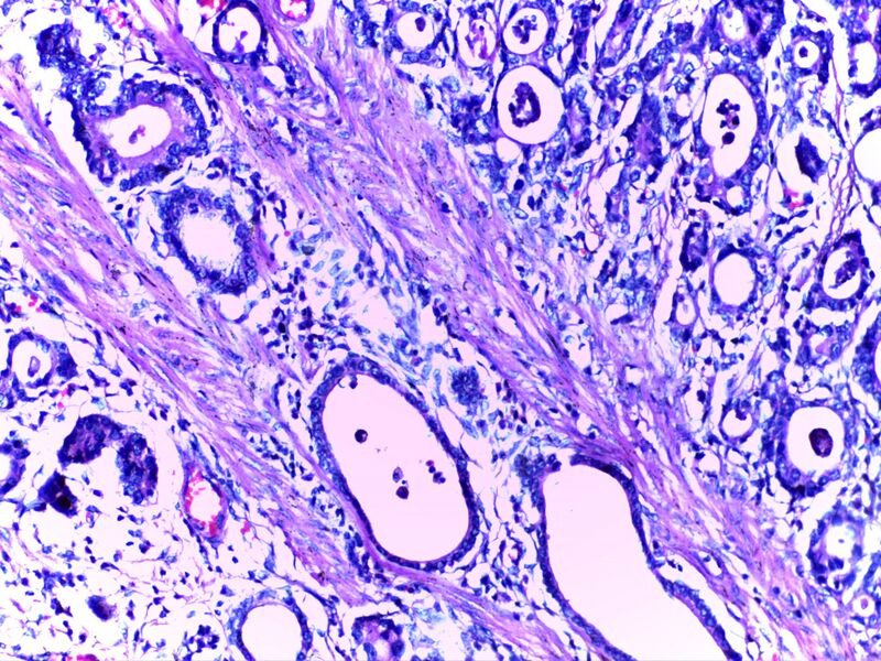
English: Micrograph of carcinoma stomach. The glands are seen infiltrating the muscle layer. The neoplastic cells are arranged in cords and in glandular pattern. The cells show dysplastic features. Signet rings with nucleus pushed to the periphery are seen in some types of adenocarcinoma.
|
| ⧼wm-license-information-date⧽ | 2015 |
| ⧼wm-license-information-source⧽ | DEPARTMENT OF PATHOLOGY, CALICUT MEDICAL COLLEGE |
| ⧼wm-license-information-author⧽ | قالب:Institution: Calicut Medical College |
ترخيص
|
|
هذا العمل مرخّص تحت رخصة المشاع الإبداعي الملزمة بنسبة العمل لمؤلفه وبترخيص الأعمال المشتقة بالمثل 4.0. |
تاريخ الملف
اضغط على زمن/تاريخ لرؤية الملف كما بدا في هذا الزمن.
| زمن/تاريخ | صورة مصغرة | الأبعاد | مستخدم | تعليق | |
|---|---|---|---|---|---|
| حالي | ★ مراجعة معتمدة 13:56، 27 أكتوبر 2023 | | 2٬048 × 1٬536 (2٫65 ميجابايت) | Pastakhov (نقاش | مساهمات) | Upload https://upload.wikimedia.org/wikipedia/commons/8/82/Carcinoma_stomach_20X.jpg |
لا يمكنك استبدال هذا الملف.
وصلات
لا يوجد صفحات تصل لهذه الصورة.