ملف:Bilirubin-from-xtal-1978-3D-balls.png
حجم هذه المعاينة: 738 × 599 بكسل. البعد الآخر: 2٬000 × 1٬624 بكسل.
الملف الأصلي (2٬000 × 1٬624 بكسل حجم الملف: 595 كيلوبايت، نوع MIME: image/png)
| ⧼wm-license-information-description⧽ |
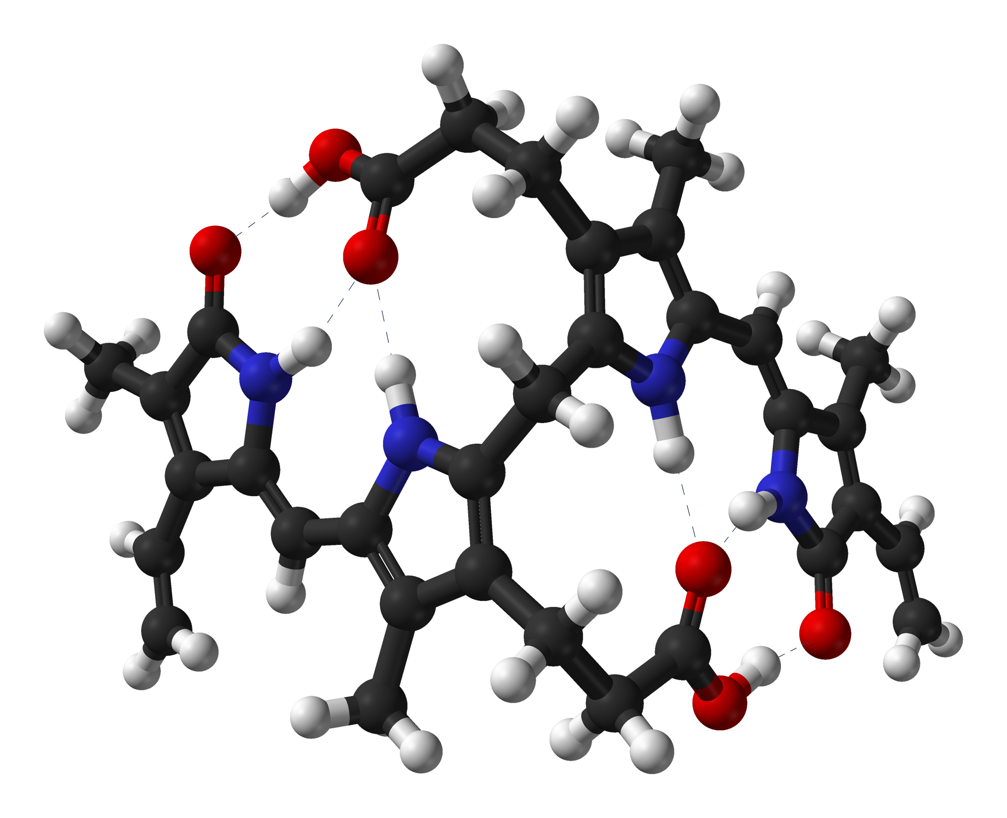
Ball-and-stick model of the bilirubin molecule as found in the crystal structure. Dashed lines are intramolecular hydrogen bonds. Colour code:
Structure by X-ray crystallography from Proc. R. Soc. Lond. B (1978) 202, 249-268. Image generated in Accelrys DS Visualizer. |
||
| ⧼wm-license-information-date⧽ | 2011 | ||
| ⧼wm-license-information-source⧽ | ⧼Wm-license-own-work⧽ | ||
| ⧼wm-license-information-author⧽ | Ben Mills | ||
| ⧼wm-license-information-permission⧽ (⧼wm-license-information-permission-reusing-text⧽) |
|
تاريخ الملف
اضغط على زمن/تاريخ لرؤية الملف كما بدا في هذا الزمن.
| زمن/تاريخ | صورة مصغرة | الأبعاد | مستخدم | تعليق | |
|---|---|---|---|---|---|
| حالي | ★ مراجعة معتمدة 03:05، 5 ديسمبر 2023 | | 2٬000 × 1٬624 (595 كيلوبايت) | Pastakhov (نقاش | مساهمات) | Upload https://upload.wikimedia.org/wikipedia/commons/3/3e/Bilirubin-from-xtal-1978-3D-balls.png |
لا يمكنك استبدال هذا الملف.
وصلات
لا يوجد صفحات تصل لهذه الصورة.