ملف:Baricitinib-portrait-ligand-3JW-from-PDB-xtal-4W9X-Mercury-3D-balls.png
حجم هذه المعاينة: 377 × 600 بكسل. البعد الآخر: 1٬257 × 2٬000 بكسل.
الملف الأصلي (1٬257 × 2٬000 بكسل حجم الملف: 588 كيلوبايت، نوع MIME: image/png)
وصف قصير
| ⧼wm-license-information-description⧽ |
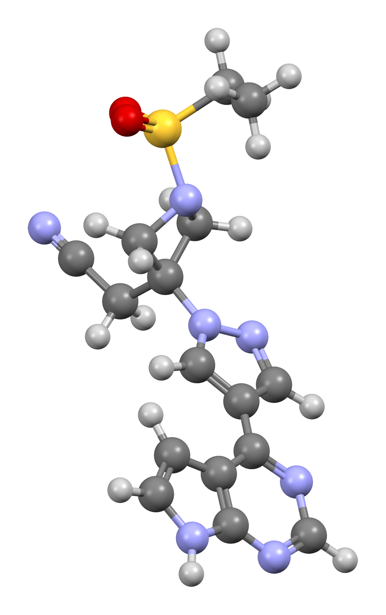
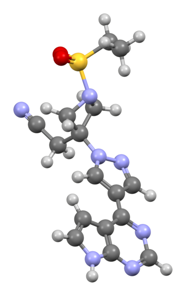
Ball-and-stick model of a baricitinib molecule, C16H17N7O2S as found in the crystal structure of BMP-2-inducible kinase in complex with baricitinib, reported in Structure (2016) 24, 401-411 (PDB entry: 4W9X; PDB ligand entry: 3JW; PDBe ligand entry: 3JW). Molecule oriented to correspond to File:Baricitinib_structure.svg as closely as possible. Colour code:
Model manipulated and image generated in CCDC Mercury 3.8. |
||
| ⧼wm-license-information-date⧽ | 2020 | ||
| ⧼wm-license-information-source⧽ | ⧼Wm-license-own-work⧽ | ||
| ⧼wm-license-information-author⧽ | Ben Mills | ||
| ⧼wm-license-information-permission⧽ (⧼wm-license-information-permission-reusing-text⧽) |
|
تاريخ الملف
اضغط على زمن/تاريخ لرؤية الملف كما بدا في هذا الزمن.
| زمن/تاريخ | صورة مصغرة | الأبعاد | مستخدم | تعليق | |
|---|---|---|---|---|---|
| حالي | ★ مراجعة معتمدة 12:26، 27 أكتوبر 2023 | | 1٬257 × 2٬000 (588 كيلوبايت) | Pastakhov (نقاش | مساهمات) | Upload https://upload.wikimedia.org/wikipedia/commons/9/94/Baricitinib-portrait-ligand-3JW-from-PDB-xtal-4W9X-Mercury-3D-balls.png |
لا يمكنك استبدال هذا الملف.
وصلات
لا يوجد صفحات تصل لهذه الصورة.