English:
Identifier: anatomydescripti1887gray (find matches)
Title: Anatomy, descriptive and surgical
Year: 1887 (1880s)
Authors: Gray, Henry, 1825-1861 Pick, T. Pickering (Thomas Pickering), 1841-1919, ed Keen, William W. (William Williams), b. 1837
Subjects: Human anatomy Anatomy
Publisher: Philadelphia : Lea brothers & co.
Contributing Library: Francis A. Countway Library of Medicine
Digitizing Sponsor: Open Knowledge Commons and Harvard Medical School
View Book Page: Book Viewer
About This Book: Catalog Entry
View All Images: All Images From Book
Click here to view book online to see this illustration in context in a browseable online version of this book.
Text Appearing Before Image:
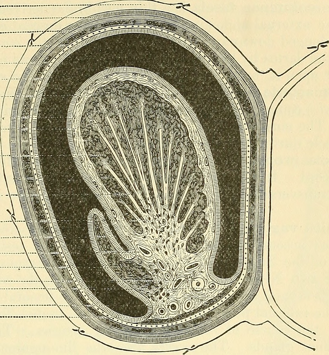
taneous nerves have Pacinian bodiesconnected with them, and, according to Krause, many of them terminate in a pecu-liar form of end-bulb. The Testes and their Coverings (Fig. 647). The testes are two small glandular organs which secrete the semen; they aresituated in the scrotum, being suspended by the spermatic cords. At an earlyperiod of foetal life the testes are contained in the abdominal cavity behind the THE PENIS. 967 peritoneum. Before birth they descend to the inguinal canal, along which theypass with the spermatic cord, and, emerging at the external abdominal ring, theydescend into the scrotum, becoming invested in their course by numerous coverings Fig. 647. Skin Dartos ■ Ext. spermatic fascia Cremasteric fascia ■ Infundibuliform fascia Parietal tunica vaginalis Visceral tunica vaginalis. Tunica albugineaA lobule of the testicle A septum MediastinumDigital fossaSpermatic veinEpididymisVas deferensArtery to vas ■ Spermatic arteryInternal muscular \ .tunic of Kolliker j
Text Appearing After Image:
Transverse Section through the Left Side of the Scrotum and the Left Testicle, the sac of the tunica vaginalisrepresented in a distended condition (Delepine). derived from the serous, muscular, and fibrous layers of the abdominal parietes, aswell as by the scrotum. The coverings of the testis are the Skin ) Q -r. V fecrotum. Dartos j Intercolumnar or External spermatic fascia. Cremasteric fascia. Infundibuliform or Fascia propria (Internal spermatic fascia). Tunica vaginalis. The Scrotum is a cutaneous pouch which contains the testes and part of thespermatic cords. It is divided into two lateral halves by a median line or raphewhich is continued forward to the under surface of the penis and backward alongthe middle line of the perineum to the anus. Of these two lateral portions, the leftis longer than the right, and corresponds with the greater length of the spermaticcord on the left side. Its external aspect varies under different circumstances:thus, under the influence of warmth and
Note About Images
Please note that these images are extracted from scanned page images that may have been digitally enhanced for readability - coloration and appearance of these illustrations may not perfectly resemble the original work.
_(14763702364).jpg&oldid=3220448)