ملف:A computed tomography brain scan showing bilateral basal ganglia calcification.jpg
حجم هذه المعاينة: 800 × 589 بكسل. البعد الآخر: 1٬200 × 883 بكسل.
الملف الأصلي (1٬200 × 883 بكسل حجم الملف: 153 كيلوبايت، نوع MIME: image/jpeg)
| ⧼wm-license-information-description⧽ |
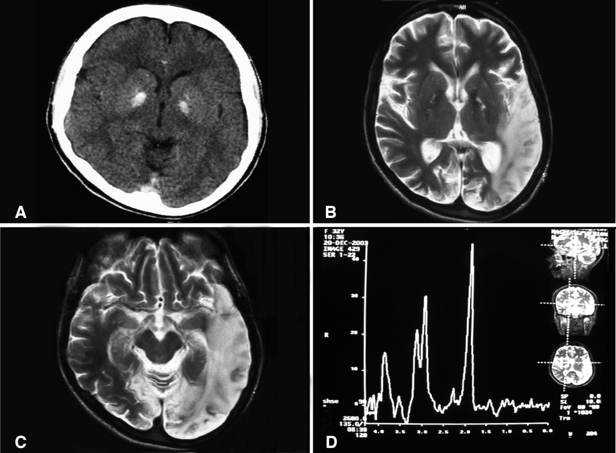
English: (a) A computed tomography brain scan showing bilateral basal ganglia calcification; the cerebellum shows prominent folia indicating mild cerebellar atrophy. (b) Axial T2 brain magnetic resonance image scan showing left temporo-parieto occipital ischemic lesion. (c) Axial T2 brain magnetic resonance image scan showing the extension of the parietal temporal region to the occipital lobe, and also showing a right occipital lesion. (d) Magnetic resonance spectroscopy showing inversion of J-coupling phenomenon at 1.3 ppm, indicating lactate peak. Abu-Amero et al. Journal of Medical Case Reports 2009 3:77 doi:10.1186/1752-1947-3-77
|
| ⧼wm-license-information-date⧽ | 2009 |
| ⧼wm-license-information-source⧽ | A patient with typical clinical features of mitochondrial encephalopathy, lactic acidosis and stroke-like episodes (MELAS) but without an obvious genetic cause: a case report |
| ⧼wm-license-information-author⧽ | Abu-Amero KK, Al-Dhalaan H, Bohlega S, Hellani A, Taylor RW. |
| ⧼wm-license-information-permission⧽ (⧼wm-license-information-permission-reusing-text⧽) |
© 2009 Abu-Amero et al; licensee BioMed Central Ltd. This is an Open Access article distributed under the terms of the Creative Commons Attribution License (https://creativecommons.org/licenses/by/2.0), which permits unrestricted use, distribution, and reproduction in any medium, provided the original work is properly cited. |
تاريخ الملف
اضغط على زمن/تاريخ لرؤية الملف كما بدا في هذا الزمن.
| زمن/تاريخ | صورة مصغرة | الأبعاد | مستخدم | تعليق | |
|---|---|---|---|---|---|
| حالي | ★ مراجعة معتمدة 00:58، 25 ديسمبر 2023 | | 1٬200 × 883 (153 كيلوبايت) | Pastakhov (نقاش | مساهمات) | Upload https://upload.wikimedia.org/wikipedia/commons/4/42/A_computed_tomography_brain_scan_showing_bilateral_basal_ganglia_calcification.jpg |
لا يمكنك استبدال هذا الملف.
وصلات
لا يوجد صفحات تصل لهذه الصورة.