ملف:3puf highlight subunit C.png
حجم هذه المعاينة: 400 × 600 بكسل.
الملف الأصلي (800 × 1٬200 بكسل حجم الملف: 379 كيلوبايت، نوع MIME: image/png)
وصف قصير
| ⧼wm-license-information-description⧽ |
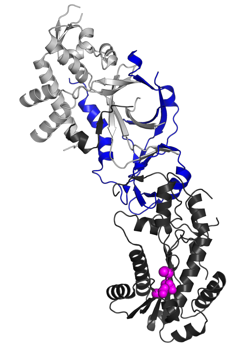
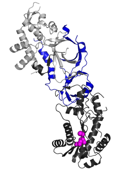
English: The human ribonuclease H2 complex. The structural subunit RNASEH2C is highlighted in blue, the catalytic subunit RNASEH2A is in dark gray, and the structural subunit RNASEH2B is in light gray. The position of the catalytic residues in the A subunit is indicated with magenta spheres.
Rendered from PDB 3PUF chains A, B, C, described in the paper: The structural and biochemical characterization of human RNase H2 complex reveals the molecular basis for substrate recognition and Aicardi-Goutieres syndrome defects. Figiel, M., Chon, H., Cerritelli, S.M., Cybulska, M., Crouch, R.J., Nowotny, M. (2011) J.Biol.Chem. 286: 10540-10550 PubMed: 21177858 PubMedCentral: PMC3060507 DOI: 10.1074/jbc.M110.181974 |
| ⧼wm-license-information-date⧽ | 2017 |
| ⧼wm-license-information-source⧽ | ⧼Wm-license-own-work⧽ |
| ⧼wm-license-information-author⧽ | Opabinia regalis |
ترخيص
|
تاريخ الملف
اضغط على زمن/تاريخ لرؤية الملف كما بدا في هذا الزمن.
| زمن/تاريخ | صورة مصغرة | الأبعاد | مستخدم | تعليق | |
|---|---|---|---|---|---|
| حالي | ★ مراجعة معتمدة 00:43، 17 أكتوبر 2023 | | 800 × 1٬200 (379 كيلوبايت) | Pastakhov (نقاش | مساهمات) | Upload https://upload.wikimedia.org/wikipedia/commons/6/6b/3puf_highlight_subunit_C.png |
لا يمكنك استبدال هذا الملف.
وصلات
لا يوجد صفحات تصل لهذه الصورة.